
















Copyright: ©Author(s) 2026.
World J Crit Care Med. Sep 9, 2026; 15(3): 120664
Published online Sep 9, 2026. doi: 10.5492/wjccm.120664
Published online Sep 9, 2026. doi: 10.5492/wjccm.120664
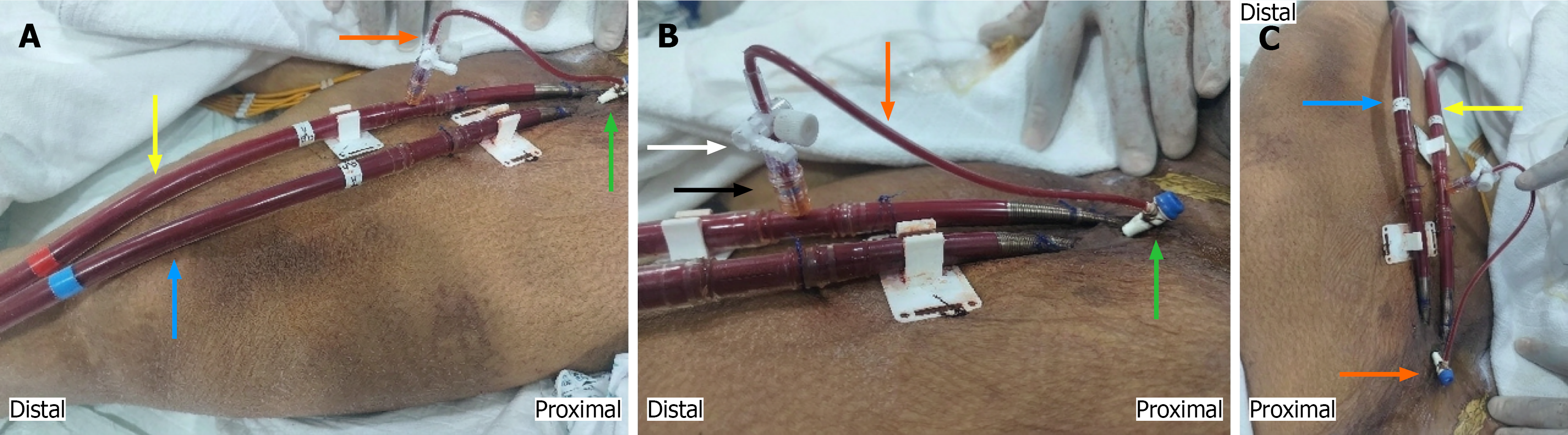
Figure 1 Image showing the distal perfusion catheter in the left lower extremity.
A: Shows distal perfusion catheter (green arrow), extension tubing with stop cock (orange arrow), venous cannula (blue arrow) and arterial cannula (yellow arrow); B: Close up view focussing on the distal perfusion catheter (green arrow) also showing connection with arterial cannula via a side port (black arrow), an intervening 3-way stop cock for regular flow check (white arrow) and a 6-inch extension tubing (orange arrow); C: Additional view showing distal perfusion catheter (orange arrow), venous cannula (blue arrow) and arterial cannula (yellow arrow).
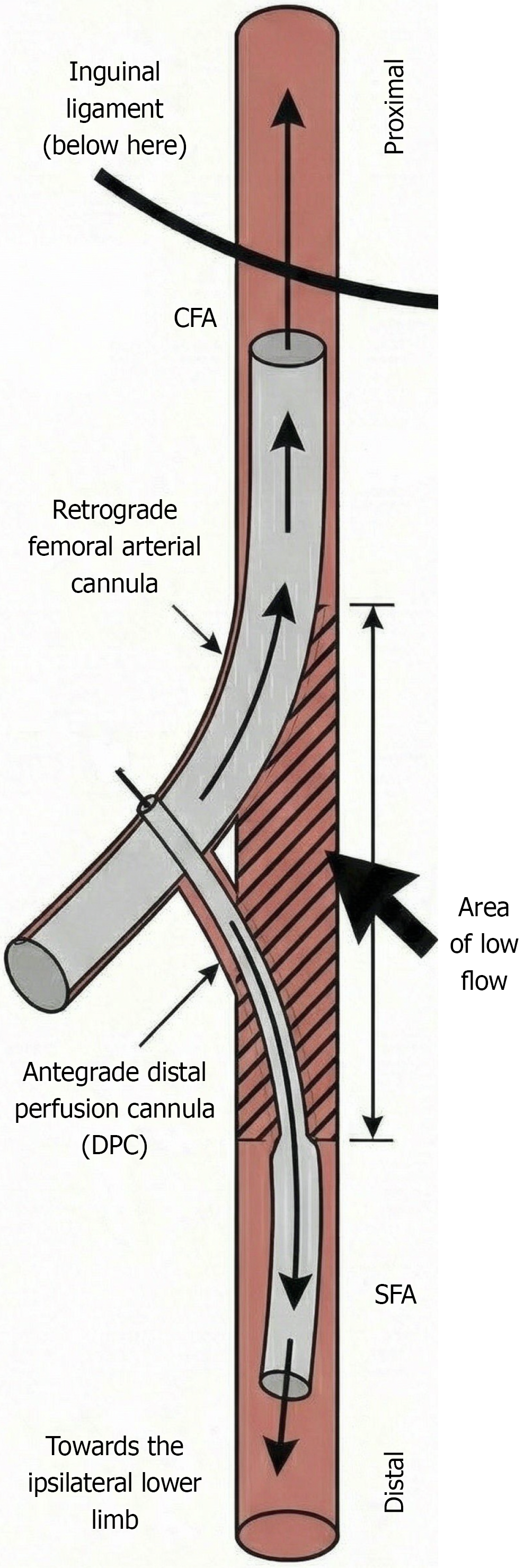
Figure 2 Illustrative image showing correct positioning and connection for placing the distal perfusion catheter.
Image shows the femoral artery with both a distal perfusion catheter and an arterial cannula inserted in opposite directions. The shaded region represents the zone where blood flow is reduced by the presence of both cannulas. SFA: Superficial femoral artery; DPC: Distal perfusion catheters; CFA: Common femoral artery.
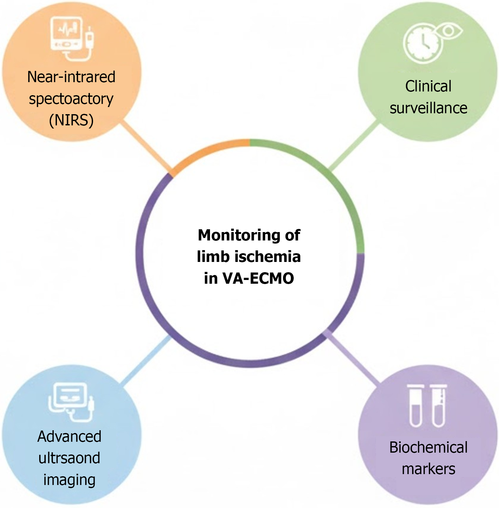
Figure 3 Multifaceted approach to monitoring limb ischemia on veno-arterial extracorporeal membrane oxygenation.
VA-ECMO: Veno-arterial extracorporeal membrane oxygenation; NIRS: Near-infrared spectroscopy.
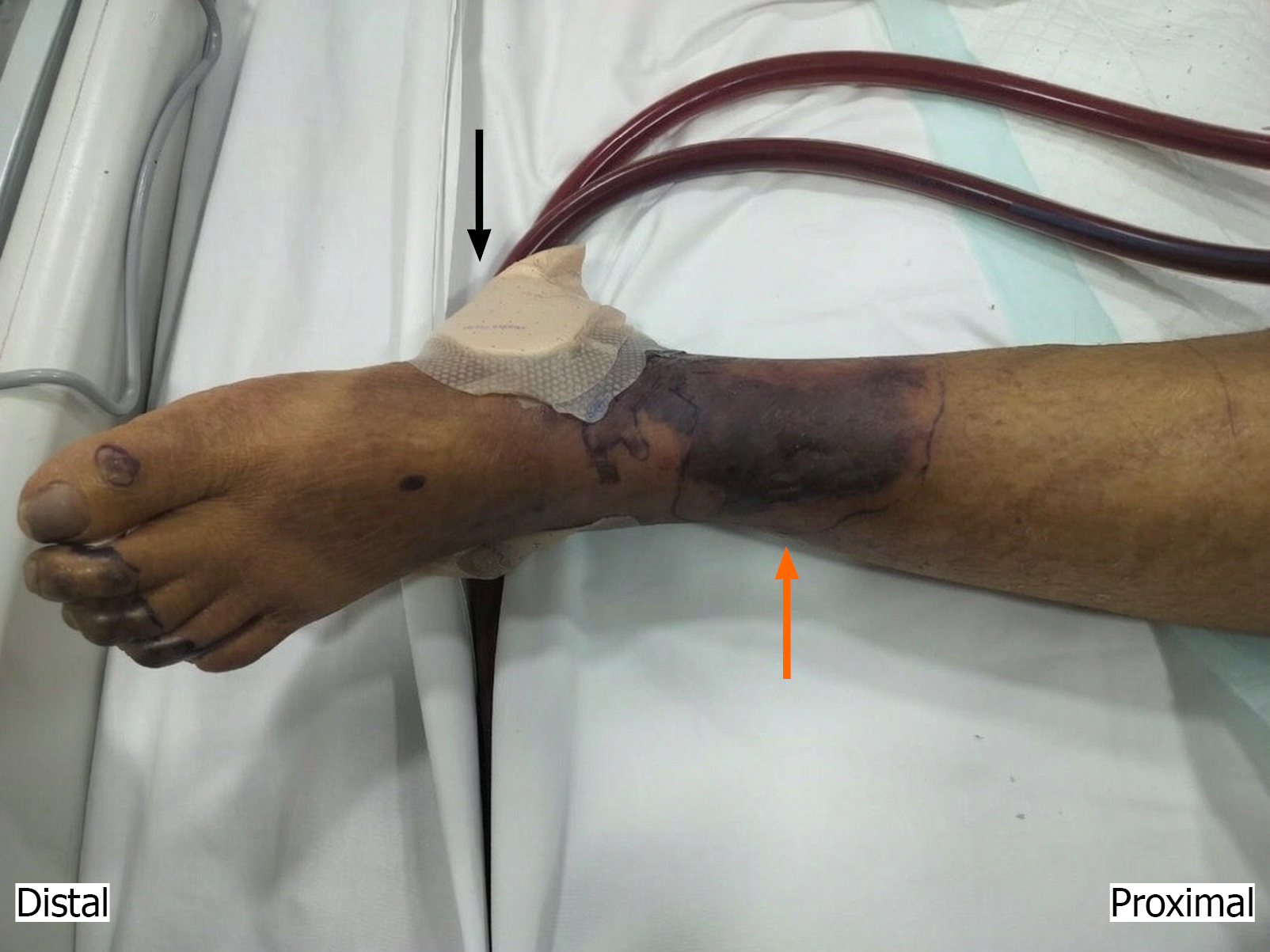
Figure 4 Left lower extremity image showing signs of limb ischemia with darkening of the skin which has been demarcated (orange arrow).
A brown coloured topical nitroglycerin patch is seen attached (black arrow).
- Citation: Karan N, Patnaik R, Pattanaik SS, Vijayakumar A, Ravinbothayan S, Behera S, Panda C, Barakat M, Meshram A, Samal S, Chawla A. Mitigating limb ischemia in veno-arterial extracorporeal membrane oxygenation: A narrative review of evolving role of distal perfusion catheters. World J Crit Care Med 2026; 15(3): 120664
- URL: https://www.wjgnet.com/2220-3141/full/v15/i3/120664.htm
- DOI: https://dx.doi.org/10.5492/wjccm.120664