
















Copyright: ©Author(s) 2026.
World J Surg Proced. Sep 16, 2026; 16(2): 122292
Published online Sep 16, 2026. doi: 10.5412/wjsp.122292
Published online Sep 16, 2026. doi: 10.5412/wjsp.122292
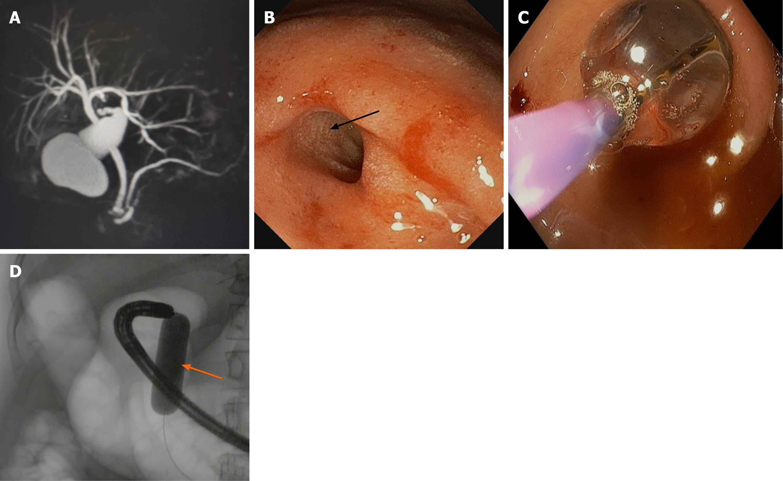
Figure 1 Preoperative investigation.
A: Magnetic resonance cholangiopancreatography showing cholelithiasis and choledocholithiasis; B: Endoscopy showing fibrotic stricture (black arrow) in the duodenum precluded a conventional endoscopic retrograde cholangiopancreatography approach; C: Endoscopy showing controlled radial expansion balloon dilatation; D: Fluoroscopy showing balloon dilatation (orange arrow) in the duodenum.
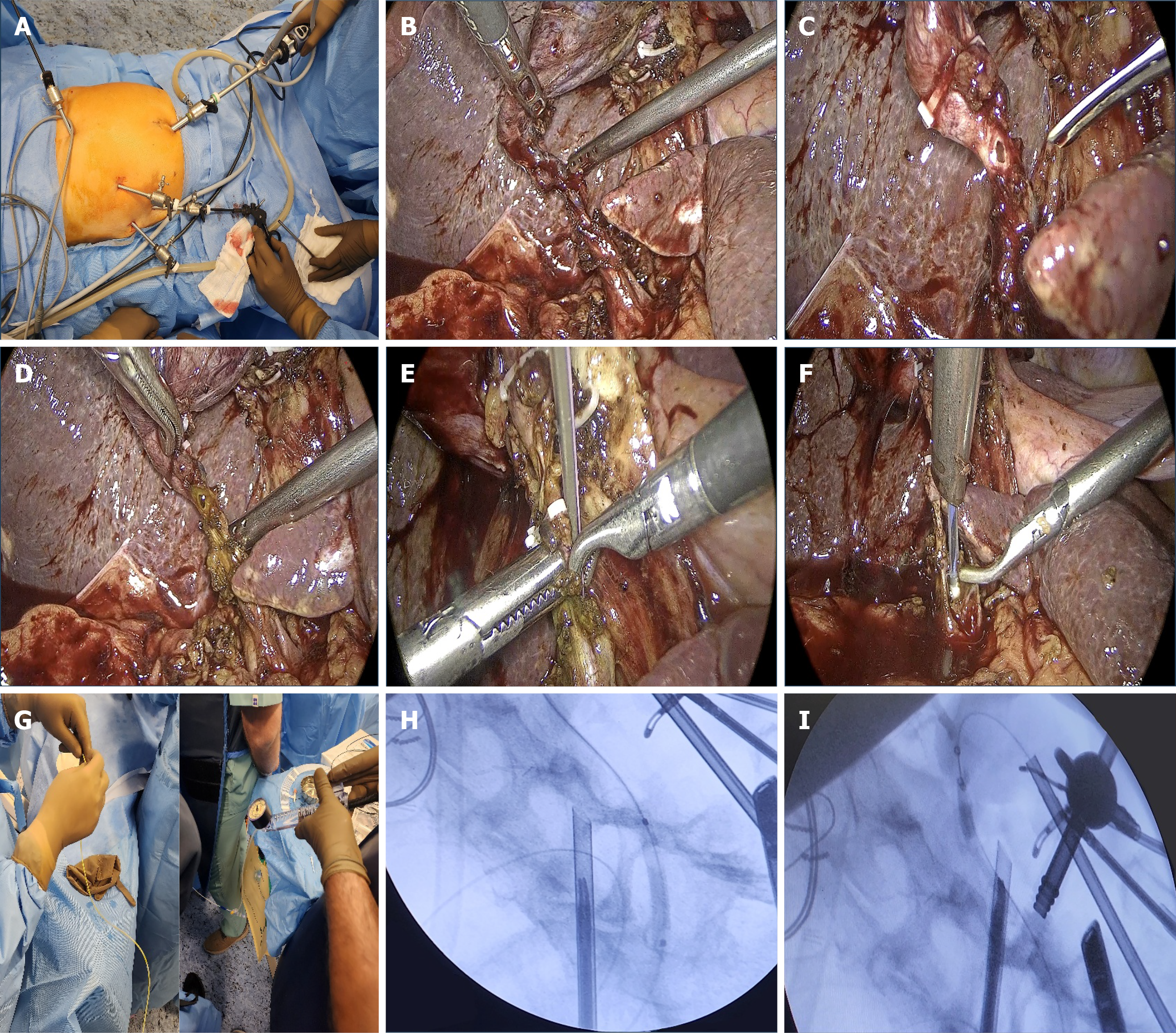
Figure 2 Intraoperative sequence of transcystic common bile duct clearance.
A: Patient and port positioning during laparoscopic cholecystectomy; B: The cystic duct stump is isolated using a grasper and suction; C: A clip is placed proximally near the junction of the cystic duct and infundibulum, before the duct is transected, showing controlled ductotomy; D: Surgical field showing bile coming out of the cystic duct opening; E and F: Transcystic approach showing a guidewire passed through a cost-effective infant feeding tube for entry into the cystic duct; G: Management of the common bile duct stone - under fluoroscopy, biliary sphincteroplasty was executed for 2 minutes with 8 atmospheres pressure; a critical step for facilitating the mechanical passage of the stone into the duodenum; H and I: Cholangiogram showing filling defect suggestive of stone and ampulla being dilated with Hurricane RX - Biliary Balloon Dilatation Catheter.
- Citation: Bhandarkar S, Dotor N, Bendapudi N, Kamat N, Parekh S, Vora S, Maydeo A. Transcystic laparoscopic common bile duct stone clearance facilitated by intraoperative fluoroscopy guided cholangiography: A case report. World J Surg Proced 2026; 16(2): 122292
- URL: https://www.wjgnet.com/2219-2832/full/v16/i2/122292.htm
- DOI: https://dx.doi.org/10.5412/wjsp.122292