
















Copyright: ©Author(s) 2026.
World J Surg Proced. Sep 16, 2026; 16(2): 118325
Published online Sep 16, 2026. doi: 10.5412/wjsp.118325
Published online Sep 16, 2026. doi: 10.5412/wjsp.118325
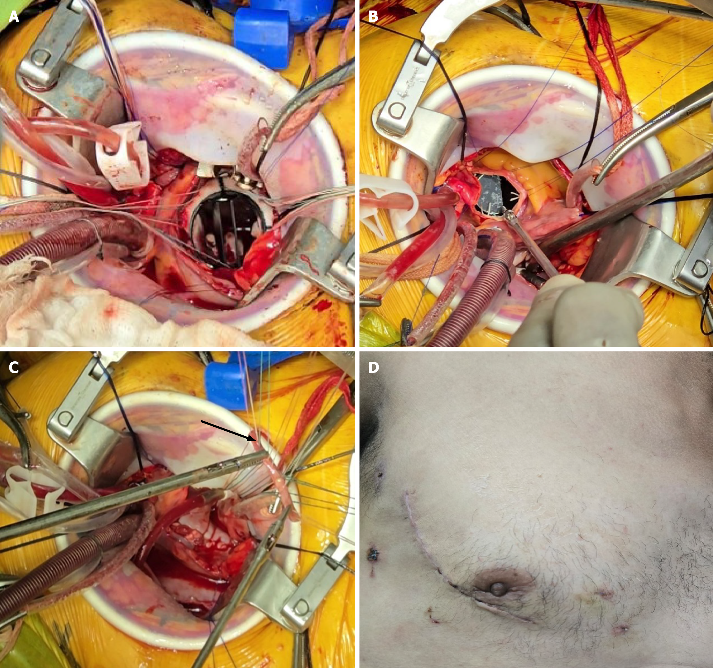
Figure 1 Intraoperative photographs.
A: Exposure of the mitral valve via intra-atrial septal approach through right atriotomy; B: Exposure of the aortic valve via transverse aortotomy; C: Tricuspid valve repair using a pericardial strip (black arrow), illustrating pericardial strip annuloplasty technique for functional tricuspid regurgitation; D: Right mini-thoracotomy incision site, highlighting the minimally invasive surgical access.
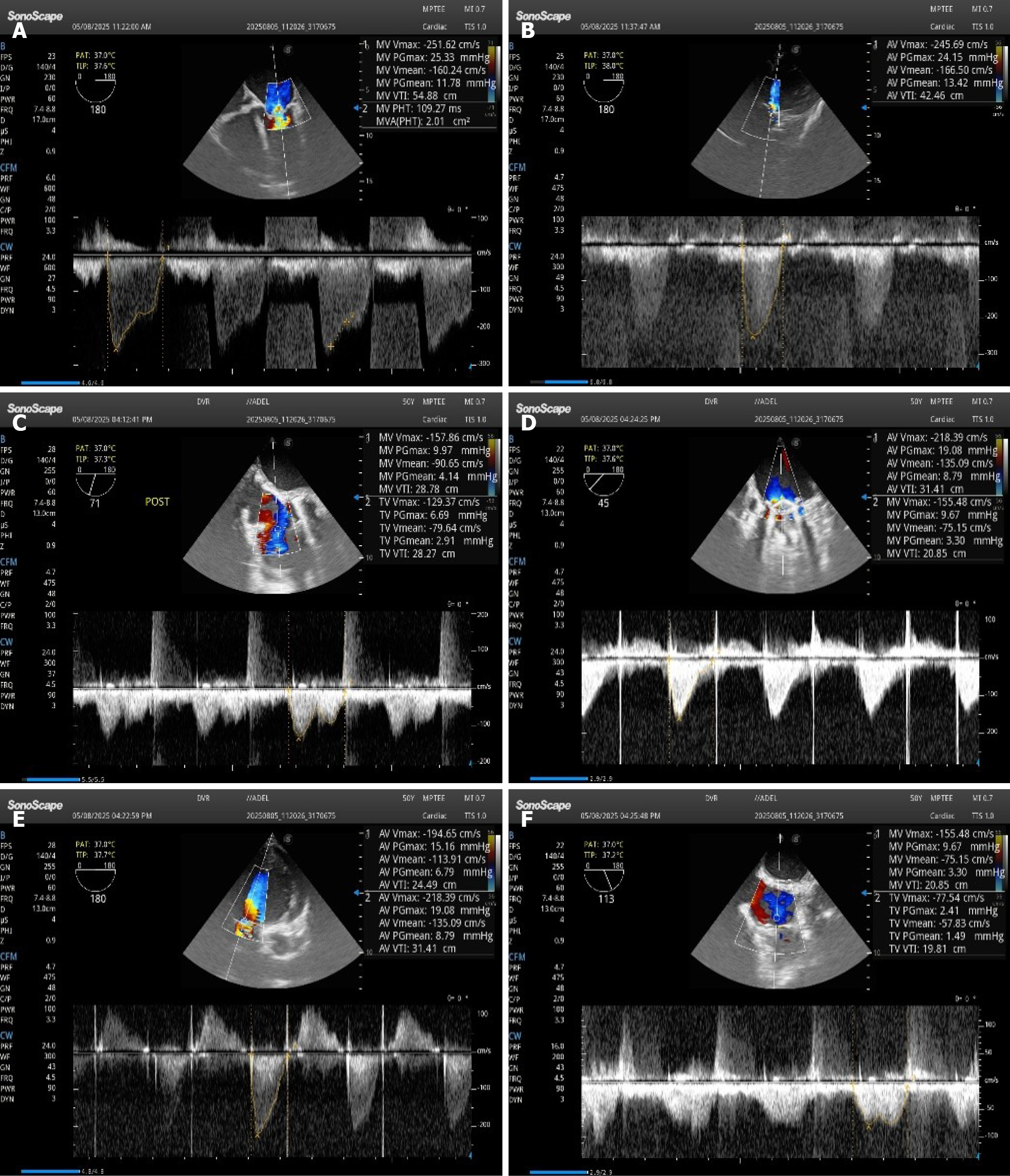
Figure 2 Echocardiographic findings.
A-C: Preoperative echocardiogram demonstrating severe multivalvular disease with restricted leaflet motion and regurgitation; D-F: Postoperative echocardiogram showing well-functioning prosthetic valves and a competent tricuspid valve, confirming successful surgical outcome.
- Citation: Al Ganadi A, Al-Shameri I, Alwsabi N, Bashraheel S, Alwsabi N, Mohammed S, Al-Fakih W. Initial experience of minimally invasive triple valve surgery in a developing country. World J Surg Proced 2026; 16(2): 118325
- URL: https://www.wjgnet.com/2219-2832/full/v16/i2/118325.htm
- DOI: https://dx.doi.org/10.5412/wjsp.118325