Copyright
©The Author(s) 2015.
World J Immunol. Nov 27, 2015; 5(3): 113-130
Published online Nov 27, 2015. doi: 10.5411/wji.v5.i3.113
Published online Nov 27, 2015. doi: 10.5411/wji.v5.i3.113
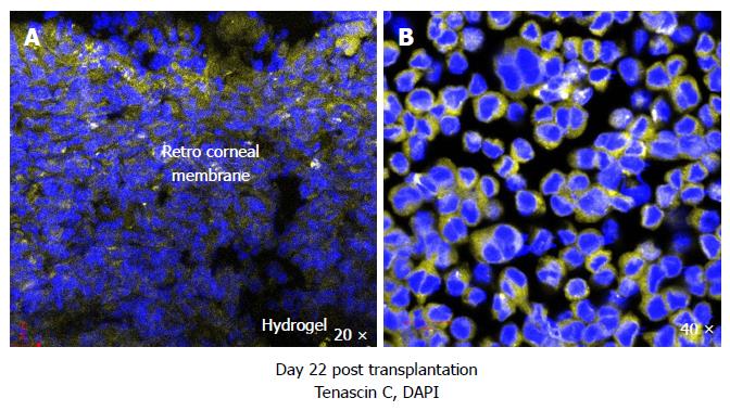
Figure 6 Murine corneas transplanted with RHCIII hydrogels stained positive for extracellular matrix constituent tenascin C in the retro-corneal membrane, a marker of epithelial to mesenchymal transition, indicative of active wound healing (A) and WEHI-164 murine fibrosarcoma cell line cultured with 5 ng/mL transforming growth factor-β1 for 48 h also produced tenascin C (positive control) (B).
DAPI: Nuclear staining shown in blue; Tenascin C in yellow.
- Citation: Shankar SP, Griffith M, Forrester JV, Kuffová L. Dendritic cells and the extracellular matrix: A challenge for maintaining tolerance/homeostasis. World J Immunol 2015; 5(3): 113-130
- URL: https://www.wjgnet.com/2219-2824/full/v5/i3/113.htm
- DOI: https://dx.doi.org/10.5411/wji.v5.i3.113


