







BPG is committed to discovery and dissemination of knowledge
Review
Copyright: ©Author(s) 2026.
World J Immunol. Nov 20, 2026; 16(3): 125225
Published online Nov 20, 2026. doi: 10.5411/wji.125225
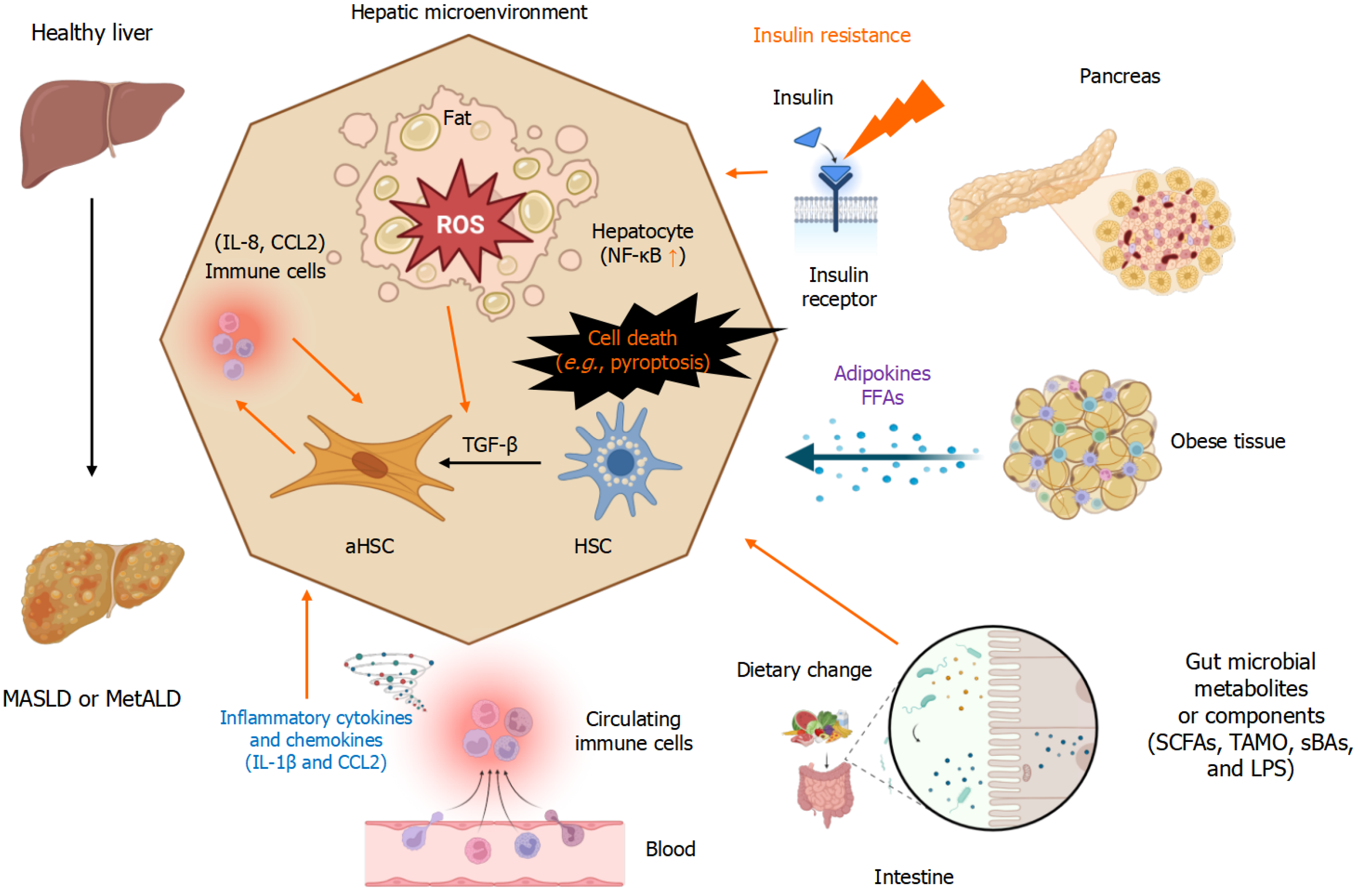
Figure 1
Figure 1 Underlying mechanisms mediating metainflammation-induced development and progression of metabolic dysfunction-associated steatotic liver disease or metabolic dysfunction and alcohol-associated liver disease. Metainflammation orchestrates the interplay among these complex interactions among insulin resistance, lipid dysregulation, oxidative stress, immune cell activation, alterations in the gut microbiota, hepatocyte injury, and fibrosis, thereby promoting the development and progression of metabolic dysfunction-associated steatotic liver disease and metabolic dysfunction and alcohol-associated liver disease. The cartoons of this figure were created using BioRender (https://www.biorender.com/). CCL2: C-C motif chemokine ligand 2; FFAs: Free fatty acids; IL-1β: Interleukin-1β; LPS: Lipopolysaccharide; MASLD: Metabolic dysfunction-associated steatotic liver disease; MetALD: Metabolic dysfunction and alcohol-associated liver disease; NF-κB: Nuclear factor kappa-light-chain-enhancer of activated B cells; ROS: Reactive oxygen species; sBAs: Secondary bile acids; SCFAs: Short-chain fatty acids; TMAO: Trimethylamine N-oxide.
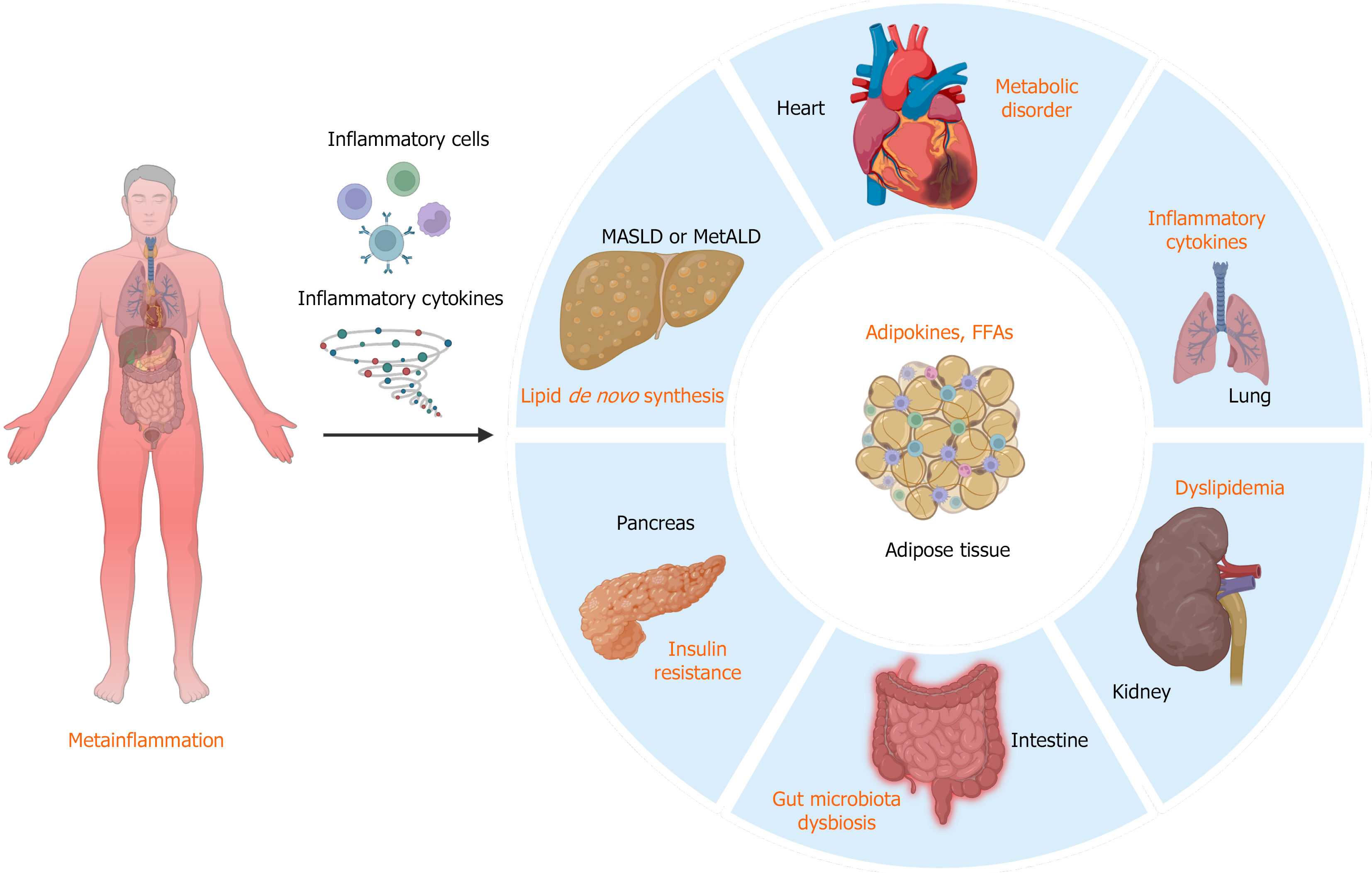
Figure 2
Figure 2 Metainflammation across different tissues and organs. The accumulation of inflammatory cells and the secretion of inflammatory cytokines facilitate interorgan communication, linking different tissues during the metabolic inflammatory state. The cartoons of this figure were created using BioRender (https://www.biorender.com/). MASLD: Metabolic dysfunction-associated steatotic liver disease; MetALD: Metabolic dysfunction and alcohol-associated liver disease; FFAs: Free fatty acids.


Write to the Help Desk