Copyright
©The Author(s) 2020.
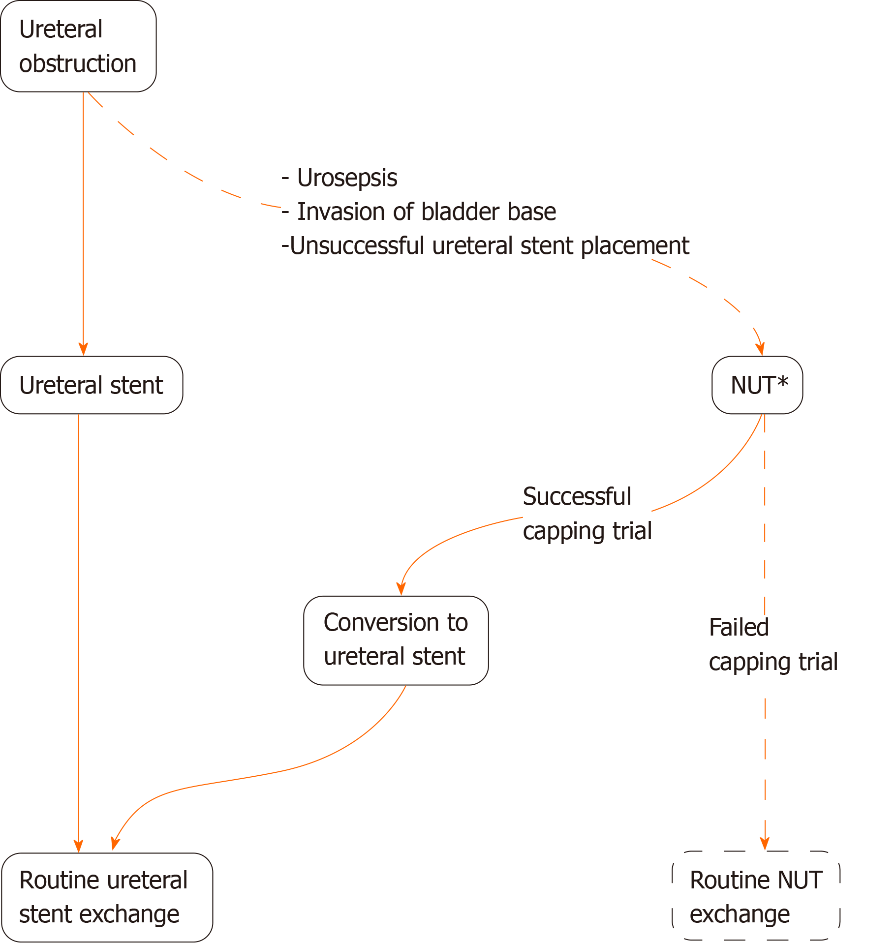
Figure 1 Algorithm of management of ureteral obstruction.
The ideal objectives are highlighted with solid lines. Study’s observation occurs when a nephroureterostomy tube is in place (asterisk). If originally a percutaneous nephrostomy catheter is placed, it will be converted to a nephroureterostomy tube before capping trial. NUT: Nephroureterostomy tube.
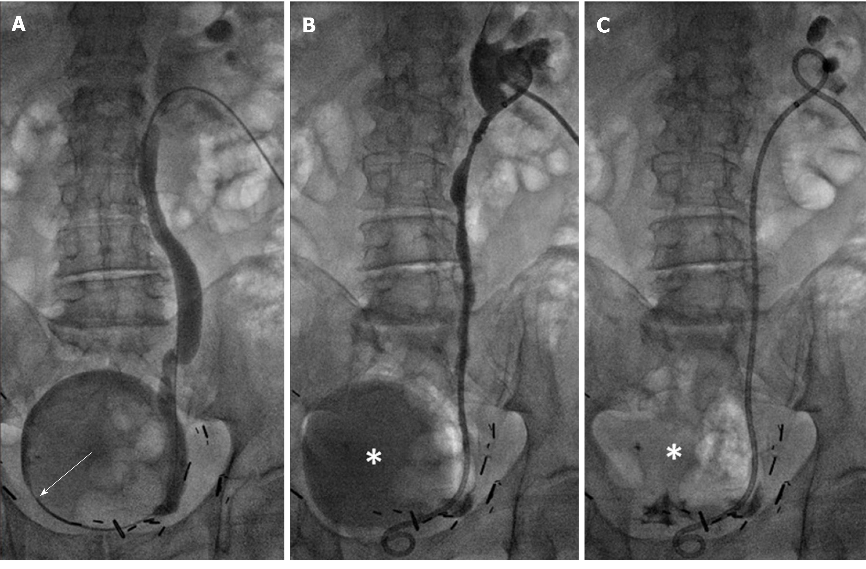
Figure 2 An 83 years old male with prostate cancer presented with right hydroureteronephrosis from a tumor involving the right bladder base.
Cystoscopic attempt at placing ureteral stent failed. The indication for drainage of kidney is to preserve renal function for chemotherapy. Prone position. A: Distal right ureteral obstruction is crossed and contrast is injected into the bladder by a 5 French directional catheter (arrow); B: A 10 F × 26 cm nephroureterostomy tube (NUT) is placed, and small amount of contrast is injected through the NUT to confirm proper positioning of the proximal loop. Retained contrast/urine in bladder (asterisk) is evident; and C: Retained contrast in bladder (asterisk) is completely aspirated via NUT. B and C are the final images of this intervention. The NUT was capped a few days later. It remained capped with no clinical issues for 42 d before it was converted to a ureteral stent.
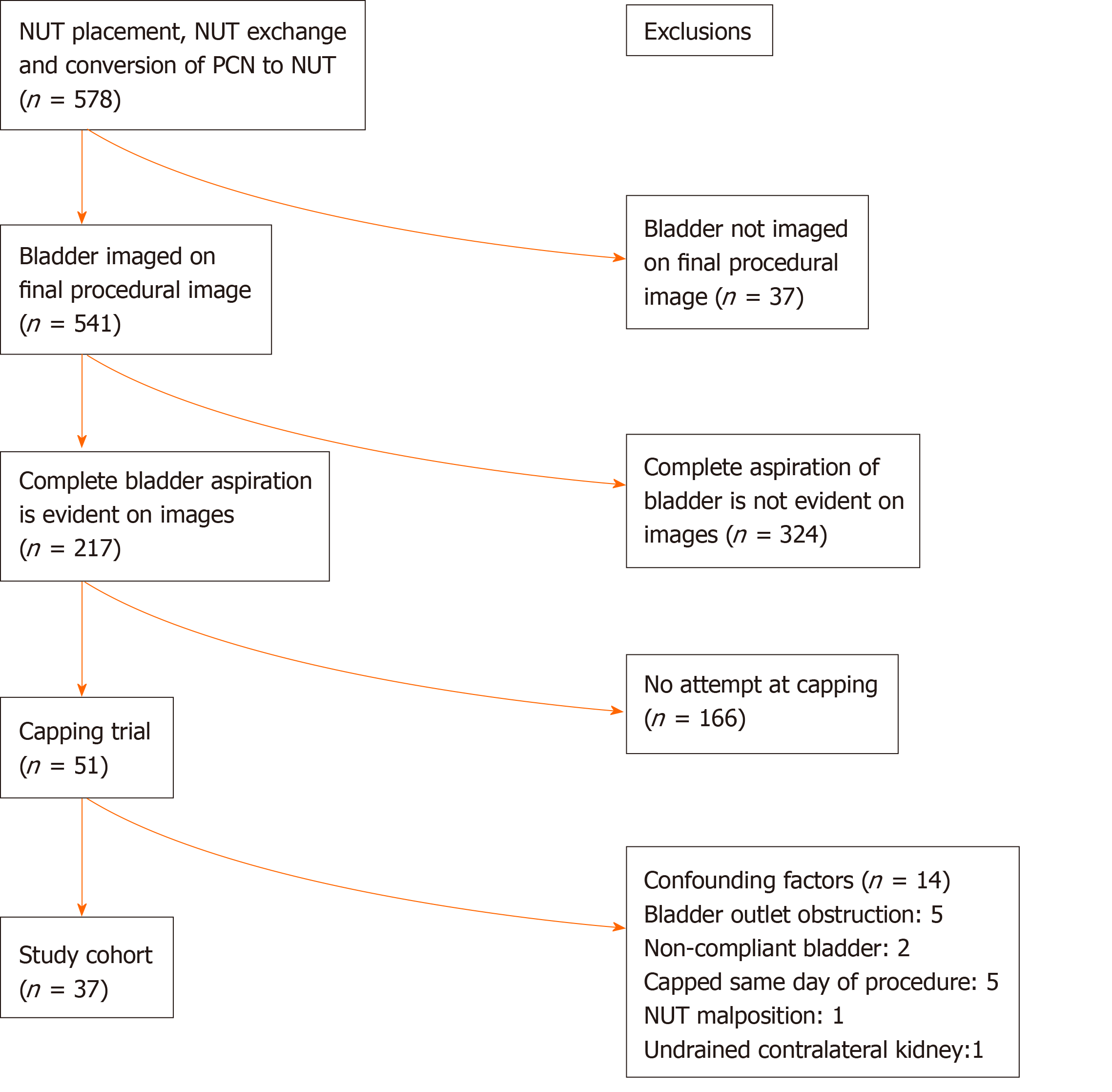
Figure 3 Flowchart of study.
NUT: Nephroureterostomy tube; PCN: Percutaneous nephrostomy tube.
- Citation: Maybody M, Shay WK, Fleischer DA, Hsu M, Moskowitz C. Estimation of successful capping with complete aspiration of bladder via nephroureterostomy tube. World J Clin Urol 2020; 9(1): 1-8
- URL: https://www.wjgnet.com/2219-2816/full/v9/i1/1.htm
- DOI: https://dx.doi.org/10.5410/wjcu.v9.i1.1


