Copyright
©The Author(s) 2016.
World J Clin Urol. Mar 24, 2016; 5(1): 72-74
Published online Mar 24, 2016. doi: 10.5410/wjcu.v5.i1.72
Published online Mar 24, 2016. doi: 10.5410/wjcu.v5.i1.72
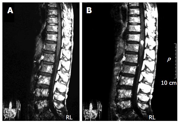
Figure 1 The thracolumbar T1-wighted magnetic resonance imaging.
Though unenhanced magnetic resonance imaging (MRI) revealed no abnormal finding (A), gadolinium diethylenetriaminepentaacetic acid enhanced MRI indicated a spot on the spinal cord at the Th12 level (B).
- Citation: Soga H, Imanishi O. Case of intramedullary spinal cord metastasis of renal cell carcinoma. World J Clin Urol 2016; 5(1): 72-74
- URL: https://www.wjgnet.com/2219-2816/full/v5/i1/72.htm
- DOI: https://dx.doi.org/10.5410/wjcu.v5.i1.72


