
















Copyright: ©Author(s) 2026.
World J Clin Urol. Sep 16, 2026; 15(2): 120632
Published online Sep 16, 2026. doi: 10.5410/wjcu.120632
Published online Sep 16, 2026. doi: 10.5410/wjcu.120632
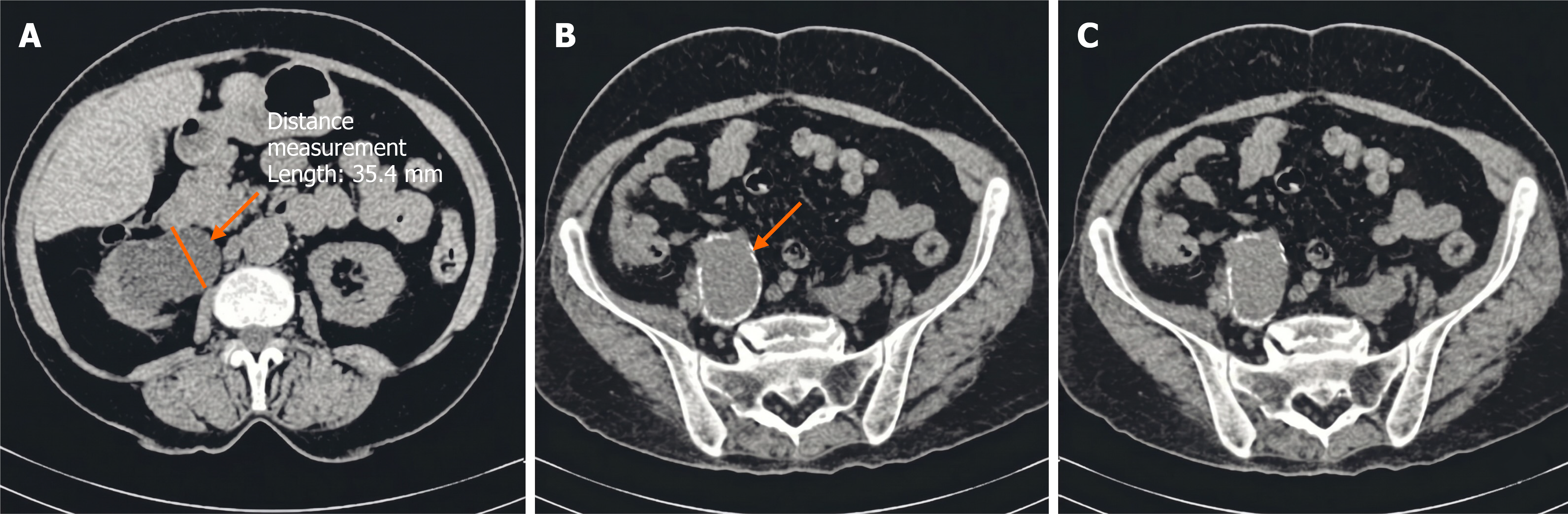
Figure 1 Noncontrast axial computed tomography images demonstrating obstructive uropathy secondary to a right common iliac artery aneurysm.
A: Right pelvicalyceal dilatation consistent with hydronephrosis (arrow); B: Right common iliac artery aneurysm with adjacent ureteral compression (arrow); C: A cut below the previous ones showing the largest part of the aneurysm. Computed tomography was performed without intravenous contrast because of acute kidney injury.
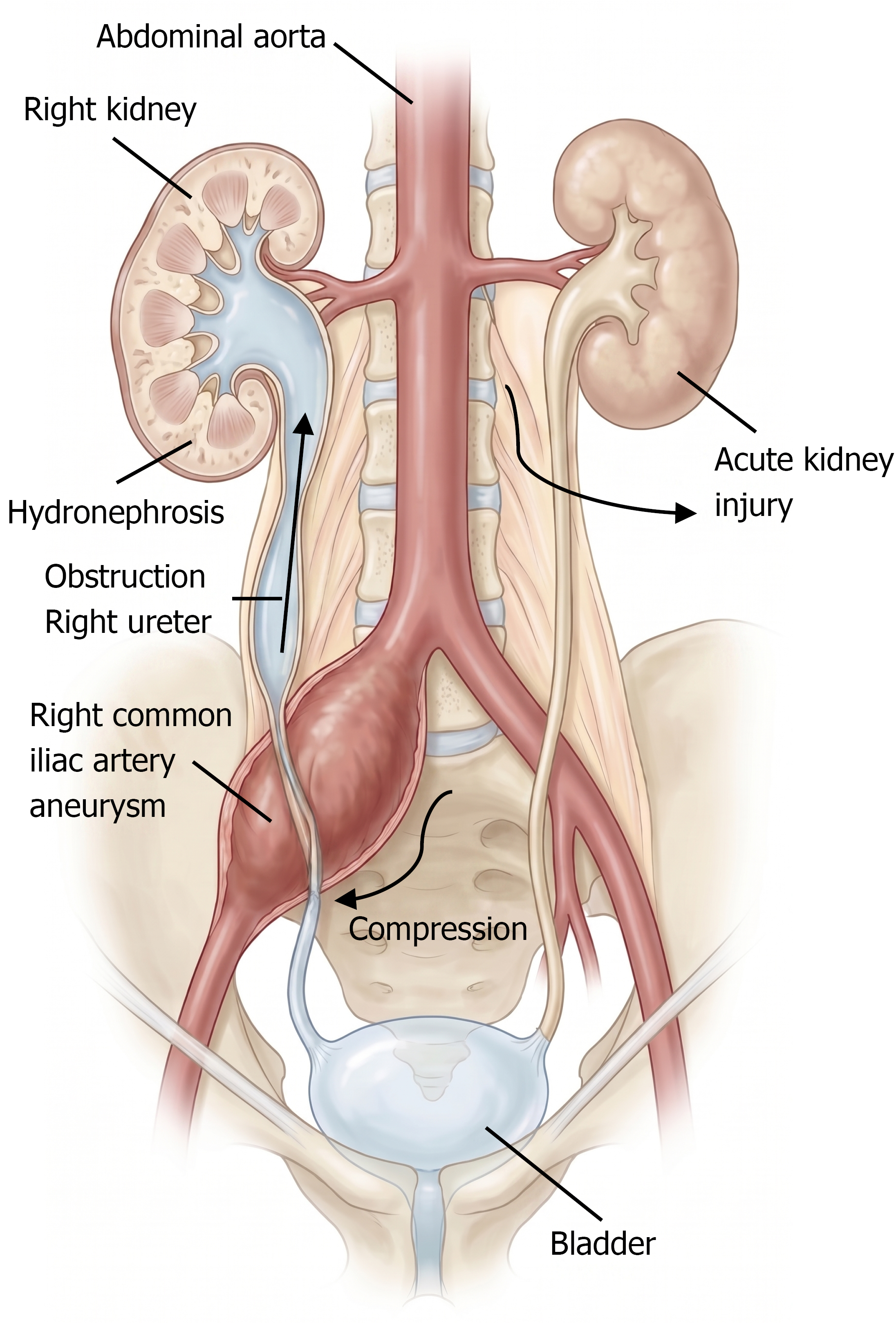
Figure 2
Schematic illustration demonstrating extrinsic compression of the right ureter by a large right common iliac artery aneurysm resulting in hydroureteronephrosis and obstructive uropathy.
- Citation: Seif S, Mahmoud S, El Sayad I, Bou Rizk R, Tlais M. Obstructive uropathy and acute-on-chronic kidney disease from common iliac artery aneurysm: A case report. World J Clin Urol 2026; 15(2): 120632
- URL: https://www.wjgnet.com/2219-2816/full/v15/i2/120632.htm
- DOI: https://dx.doi.org/10.5410/wjcu.120632