Copyright
©The Author(s) 2022.
World J Clin Urol. Aug 29, 2022; 11(1): 1-5
Published online Aug 29, 2022. doi: 10.5410/wjcu.v11.i1.1
Published online Aug 29, 2022. doi: 10.5410/wjcu.v11.i1.1
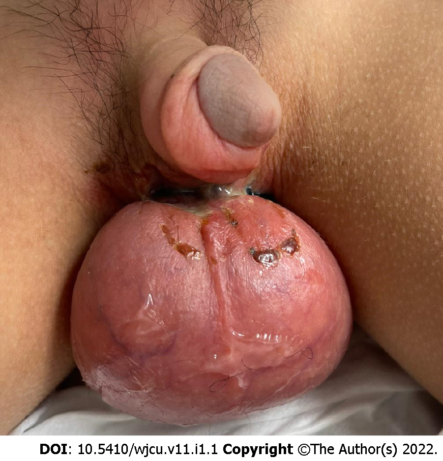
Figure 1 Bilateral scrotal redness and swelling.
Note a serous discharge on the scrotal skin, which appears edematous.
Figure 2 Scrotal ultrasound.
A: Bilateral view; B: Normal blood flow on right; C: Left testicle.
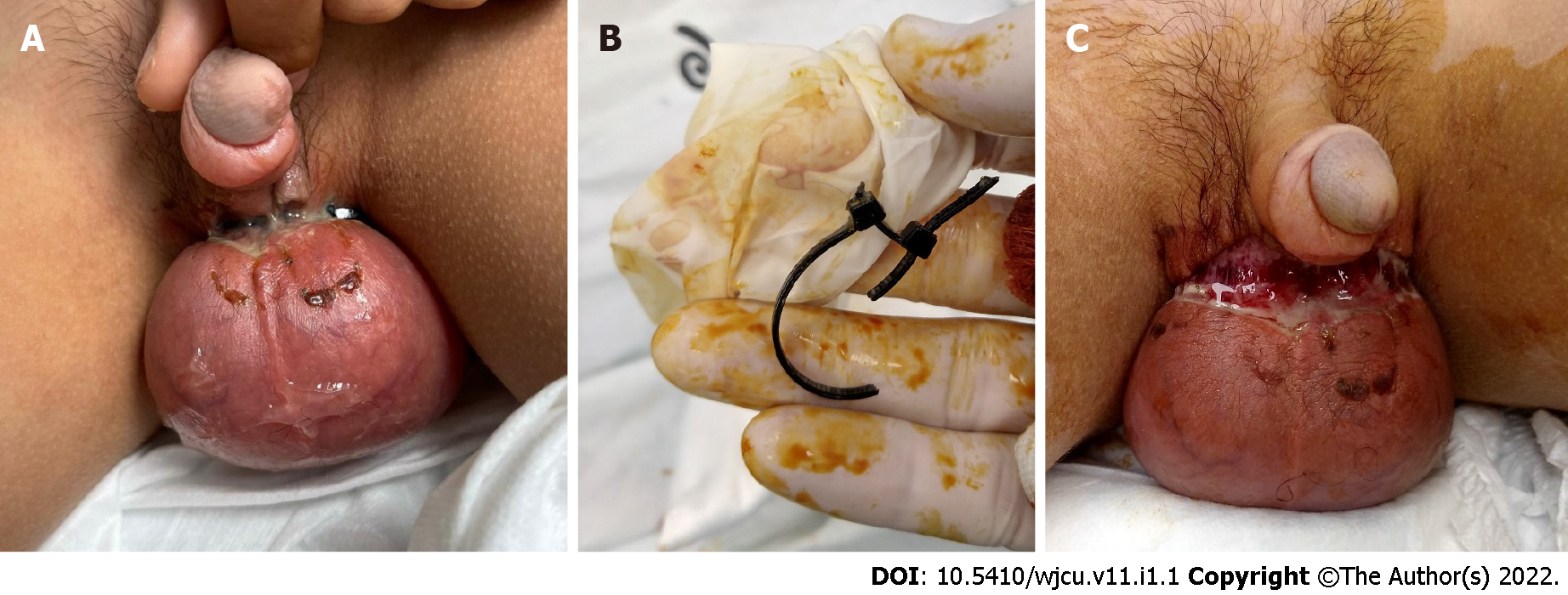
Figure 3 Idiopathic scrotal edema.
A and B: Scrotal entrapment by zip-tie; C: Friable skin with ulceration and purulent drainage.
- Citation: Frumer M, Ben-Meir D. Scrotal strangulation in the differential diagnosis of acute scrotum: A case report. World J Clin Urol 2022; 11(1): 1-5
- URL: https://www.wjgnet.com/2219-2816/full/v11/i1/1.htm
- DOI: https://dx.doi.org/10.5410/wjcu.v11.i1.1


