Copyright
©The Author(s) 2016.
World J Clin Pediatr. Aug 8, 2016; 5(3): 273-280
Published online Aug 8, 2016. doi: 10.5409/wjcp.v5.i3.273
Published online Aug 8, 2016. doi: 10.5409/wjcp.v5.i3.273
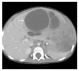
Figure 6 Mesenchymal hamartoma.
15-mo-old with abdominal distention: Axial computed tomography scan after the administration of IV contrast material demonstrates a large multi-cystic mass arising from the liver (arrow). Additional mixed solid and cystic elements are present laterally in the expanded left hepatic lobe.
- Citation: Gnarra M, Behr G, Kitajewski A, Wu JK, Anupindi SA, Shawber CJ, Zavras N, Schizas D, Salakos C, Economopoulos KP. History of the infantile hepatic hemangioma: From imaging to generating a differential diagnosis. World J Clin Pediatr 2016; 5(3): 273-280
- URL: https://www.wjgnet.com/2219-2808/full/v5/i3/273.htm
- DOI: https://dx.doi.org/10.5409/wjcp.v5.i3.273


