Copyright
©The Author(s) 2016.
World J Clin Pediatr. Aug 8, 2016; 5(3): 273-280
Published online Aug 8, 2016. doi: 10.5409/wjcp.v5.i3.273
Published online Aug 8, 2016. doi: 10.5409/wjcp.v5.i3.273
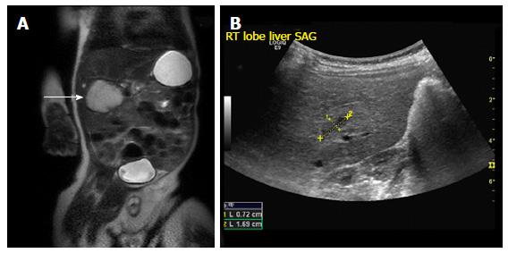
Figure 1 Focal infantile hepatic hemangiomas.
A: Coronal T2 weighted MRI image through the abdomen of an 8-wk-old boy revealing a large hyperintense mass arising from the liver (arrow); B: Abdominal USG of the same patient at 17-mo-old shows a minimal residual scar (demarcated by calipers). USG: Ultrasonography; MRI: Magnetic resonance imaging.
- Citation: Gnarra M, Behr G, Kitajewski A, Wu JK, Anupindi SA, Shawber CJ, Zavras N, Schizas D, Salakos C, Economopoulos KP. History of the infantile hepatic hemangioma: From imaging to generating a differential diagnosis. World J Clin Pediatr 2016; 5(3): 273-280
- URL: https://www.wjgnet.com/2219-2808/full/v5/i3/273.htm
- DOI: https://dx.doi.org/10.5409/wjcp.v5.i3.273


