Copyright
©2013 Baishideng Publishing Group Co.
World J Stomatol. Nov 20, 2013; 2(4): 97-102
Published online Nov 20, 2013. doi: 10.5321/wjs.v2.i4.97
Published online Nov 20, 2013. doi: 10.5321/wjs.v2.i4.97
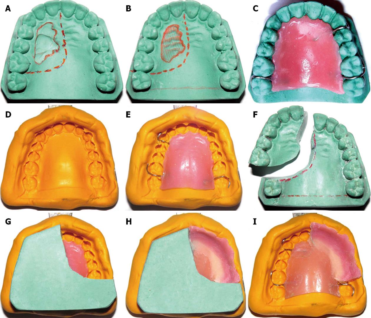
Figure 1 Technique to fabricate surgical obturator duplicating original tissue-form.
A: Working cast; B: Modification of cast; C: Prosthetic base; D: Putty impression index; E: Prosthetic base transferred onto putty index; F: Maxillary cast sectioned; G: Reseating of putty index onto sectioned cast; H: Creation of the prosthetic teeth in tooth-colored and facial surface in pink-colored resin; I: Removal of cast from index.
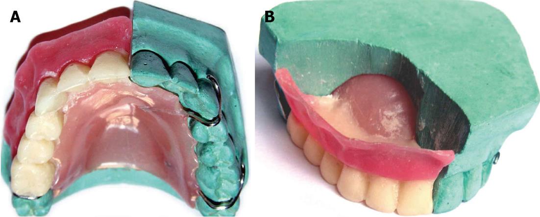
Figure 2 Completed obturator.
A: Palatal view; B: Obturator in relation with remaining normal structures on cast.
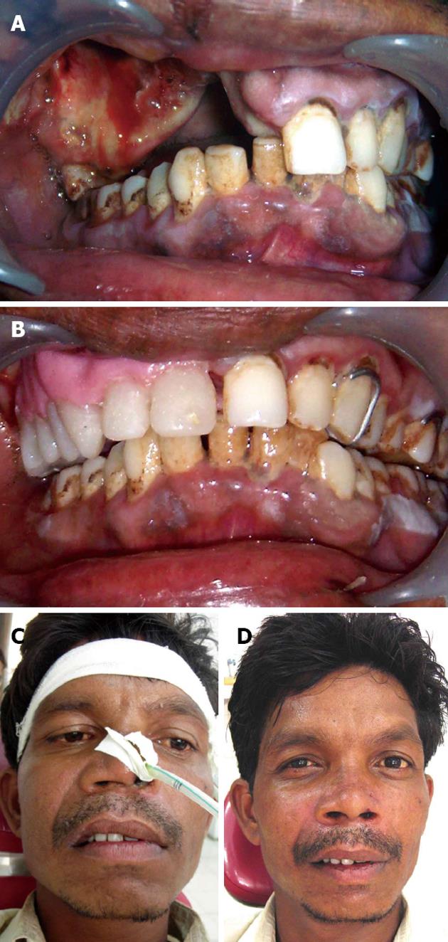
Figure 3 Post-treatment view immediate and after 6 mo.
A: Immediate post-surgical view; B: Obturator in place; C: Immediate post treatment extraoral view; D: Six month post treatment extraoral view.
- Citation: Patil PG. Surgical obturator duplicating original tissue-form restores esthetics and function in oral cancer. World J Stomatol 2013; 2(4): 97-102
- URL: https://www.wjgnet.com/2218-6263/full/v2/i4/97.htm
- DOI: https://dx.doi.org/10.5321/wjs.v2.i4.97


