Copyright
©2013 Baishideng.
World J Stomatol. Feb 20, 2013; 2(1): 12-17
Published online Feb 20, 2013. doi: 10.5321/wjs.v2.i1.12
Published online Feb 20, 2013. doi: 10.5321/wjs.v2.i1.12
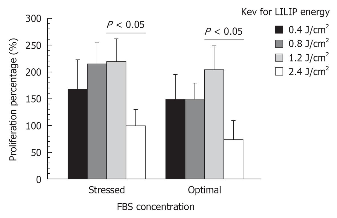
Figure 1 Bar chart of the stem cells from human exfoliated deciduous teeth proliferation within the dental pulp constructs with different low intensity laser irradiation phototherapy energy densities and different fetal bovine serum concentrations.
LILIP: Low intensity laser irradiation phototherapy; FBS: Fetal bovine serum.
Figure 2 Bar chart of the mineralization percentages of stem cells from human exfoliated deciduous teeth cultures with different low intensity laser irradiation phototherapy energy densities and different fetal bovine serum concentrations.
LILIP: Low intensity laser irradiation phototherapy; FBS: Fetal bovine serum.
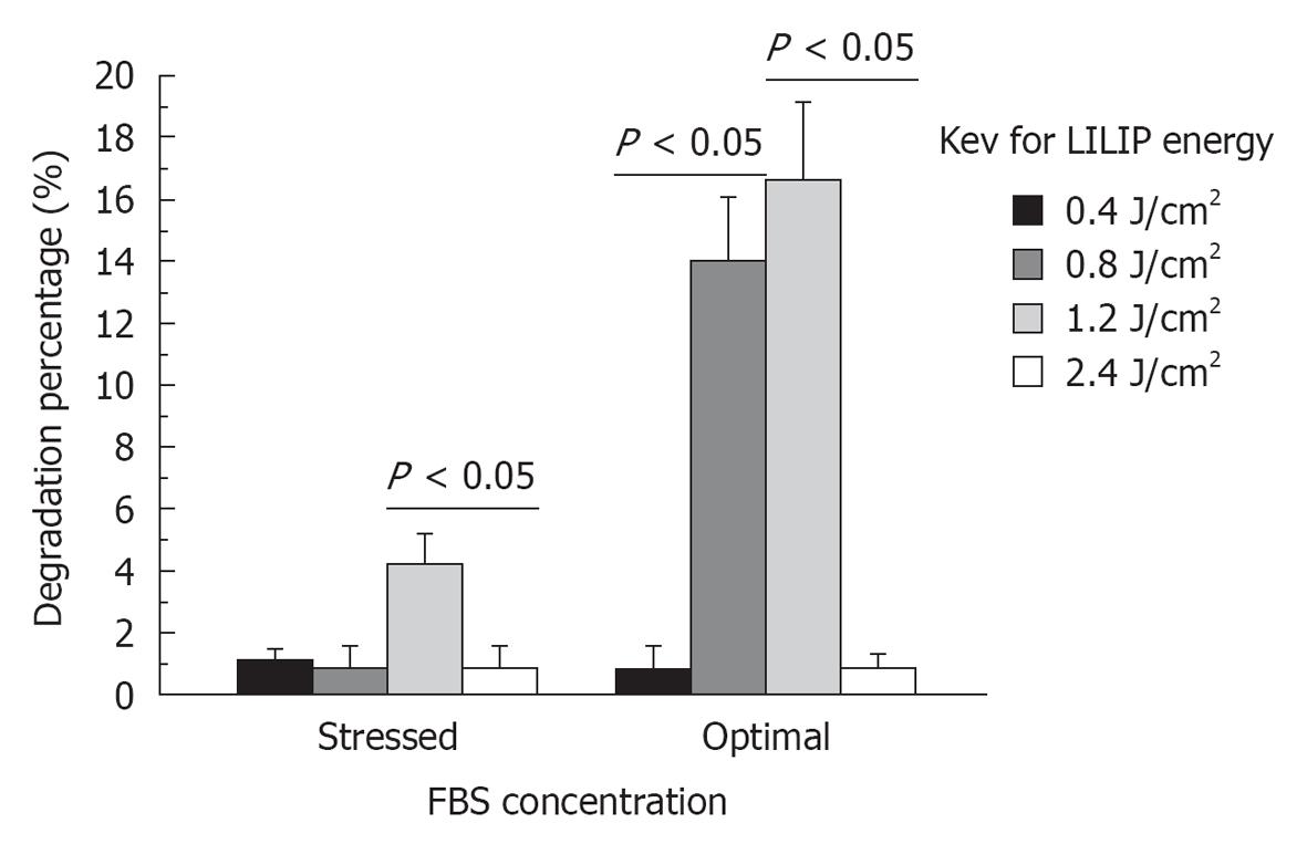
Figure 3 Bar chart of the degradation within the dental pulp constructs with different low intensity laser irradiation phototherapy energy densities and different fetal bovine serum concentrations.
LILIP: Low intensity laser irradiation phototherapy; FBS: Fetal bovine serum.
- Citation: Elnaghy AM, Murray PE, Bradley P, Marchesan M, Namerow KN, Badr AE, El-Hawary YM, Badria FA. Effects of low intensity laser irradiation phototherapy on dental pulp constructs. World J Stomatol 2013; 2(1): 12-17
- URL: https://www.wjgnet.com/2218-6263/full/v2/i1/12.htm
- DOI: https://dx.doi.org/10.5321/wjs.v2.i1.12


