Copyright
©The Author(s) 2015.
World J Respirol. Jul 28, 2015; 5(2): 176-179
Published online Jul 28, 2015. doi: 10.5320/wjr.v5.i2.176
Published online Jul 28, 2015. doi: 10.5320/wjr.v5.i2.176
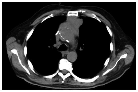
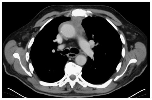
Figure 1 Computed tomography thoracic scan showing an anterior mediastinal mass with lobular contours measuring 60 mm × 66 mm.
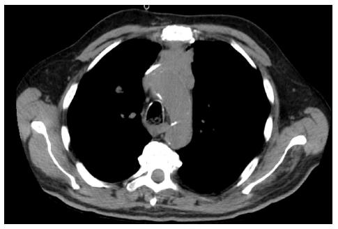
Figure 2 Computed tomography thoracic scan showing a 14-mm pulmonary nodule.
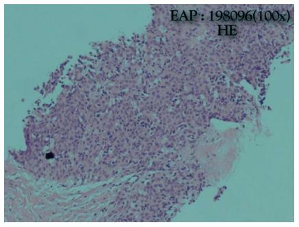
Figure 3 High-power view of poorly differentiated non-keratinizing squamous thymic carcinoma.
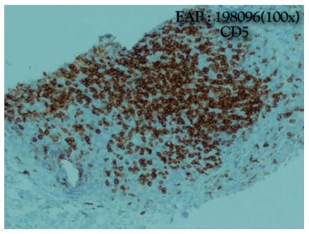
Figure 4 Malignant epithelial tumor of the thymus positive for CD5 which favors thymic carcinoma over thymoma and tumors of non-thymic origin.
A type C (World Health Organization) stage IVB (Masaoka- Koga Staging) thymic carcinoma was diagnosed.
Figure 5 Computed tomography thoracic scan showing a 27 mm × 28 mm anterior mediastinal mass.
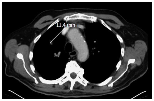
Figure 6 Computed tomography thoracic scan showing in the right superior lobe an 11-mm pulmonary nodule.
- Citation: de Macedo JE, Lopes S, Gouveia H, Oliveira S, Cunha J, Faria AL, Rego S, Oliveira A, Krug L, Bravo EM. Myasthenia gravis as a form of clinical presentation of thymic carcinoma. World J Respirol 2015; 5(2): 176-179
- URL: https://www.wjgnet.com/2218-6255/full/v5/i2/176.htm
- DOI: https://dx.doi.org/10.5320/wjr.v5.i2.176


