Copyright
©The Author(s) 2024.
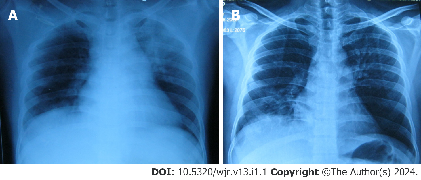
Figure 1 Chest X-ray.
A: Left upper zone non homogenous opacity with air bronchogram and right lower zone opacity; B: Resolved left upper zone opacity and non-homogenous right lower zone opacity.
Figure 2 Computed tomography pulmonary angiography.
A: Right lower lobe wedge shaped peripheral opacity suggestive of pulmonary infarct; B: Thrombus completely occluded and dilated right main pulmonary artery; C: Thrombus extending to right atrium.
- Citation: Mujeeb Rahman KK, Durgeshwar G, Mohapatra PR, Panigrahi MK, Mahanty S. Pulmonary infarct masquerading as community-acquired pneumonia in the COVID-19 scenario: A case report. World J Respirol 2024; 13(1): 1-6
- URL: https://www.wjgnet.com/2218-6255/full/v13/i1/1.htm
- DOI: https://dx.doi.org/10.5320/wjr.v13.i1.1


