Copyright
©2013 Baishideng.
World J Otorhinolaryngol. May 28, 2013; 3(2): 26-34
Published online May 28, 2013. doi: 10.5319/wjo.v3.i2.26
Published online May 28, 2013. doi: 10.5319/wjo.v3.i2.26
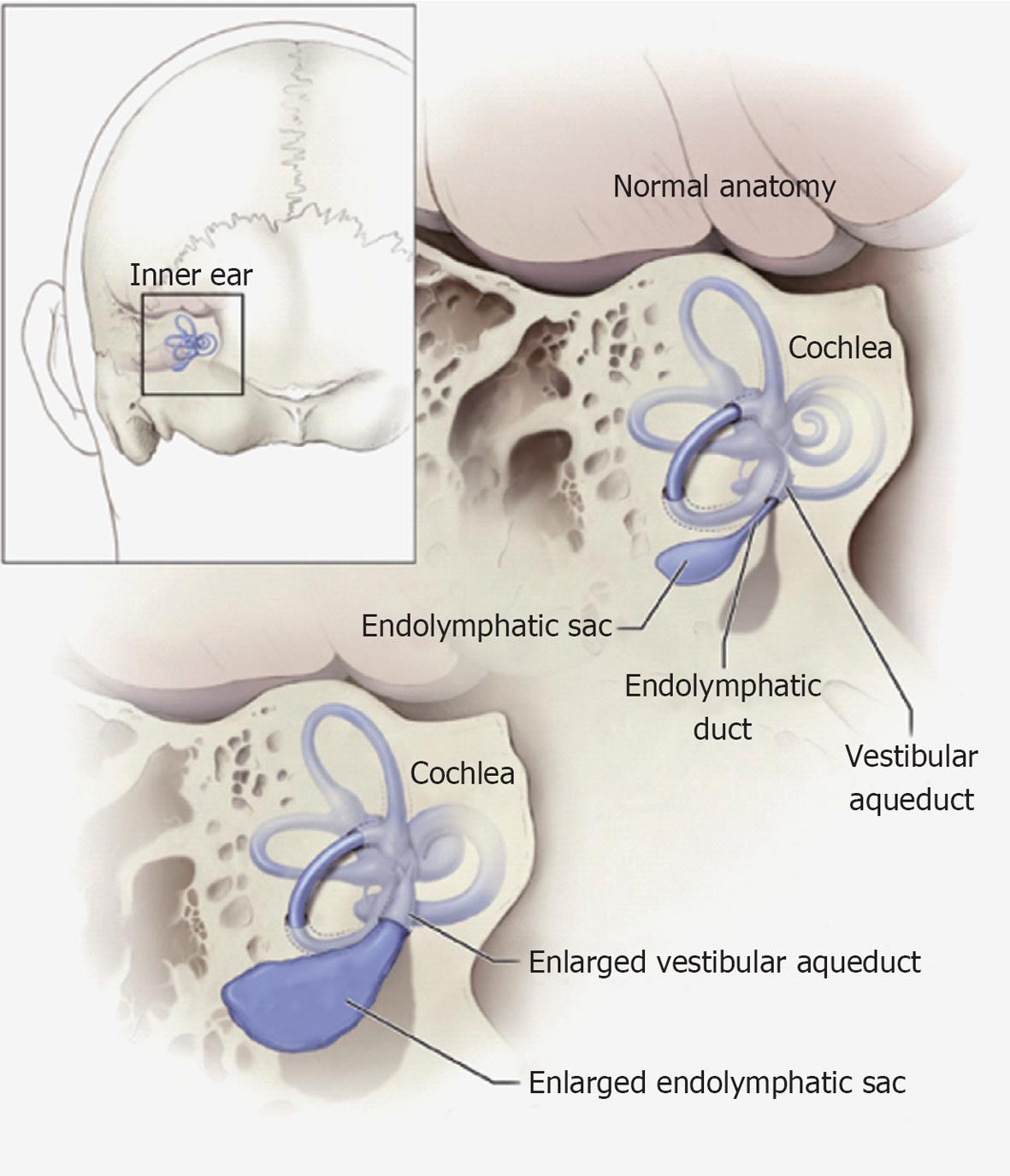
Figure 1 Schematic illustration of the relationship of the vestibular aqueduct with the endolymphatic sac and duct.
Normal anatomy of the inner ear structures is shown above. Pathologic enlargement of the endolymphatic sac and abnormal enlargement of the vestibular aqueduct are shown below. Some ears with enlargement of the vestibular aqueduct also have a reduced number of cochlear turns. Reproduced from http://www.nidcd.nih.gov/health/hearing/vestAque.htm.
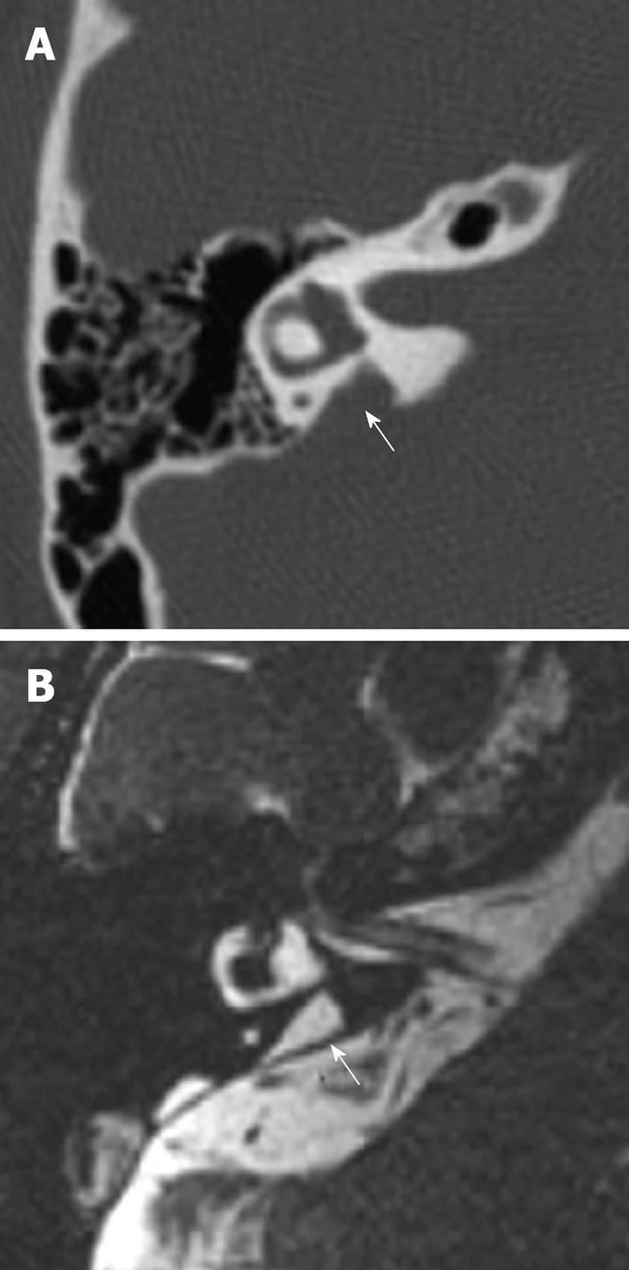
Figure 2 Right temporal bone of a patient with enlargement of the vestibular aqueduct.
A: Axial computer tomography image of a right temporal bone with an enlarged vestibular aqueduct (arrow); B: Equivalent magnetic resonance image of the same temporal bone showing an enlarged endolymphatic duct (arrow). Reproduced from http://www.nidcd.nih.gov/health/hearing/Pages/eva.aspx.
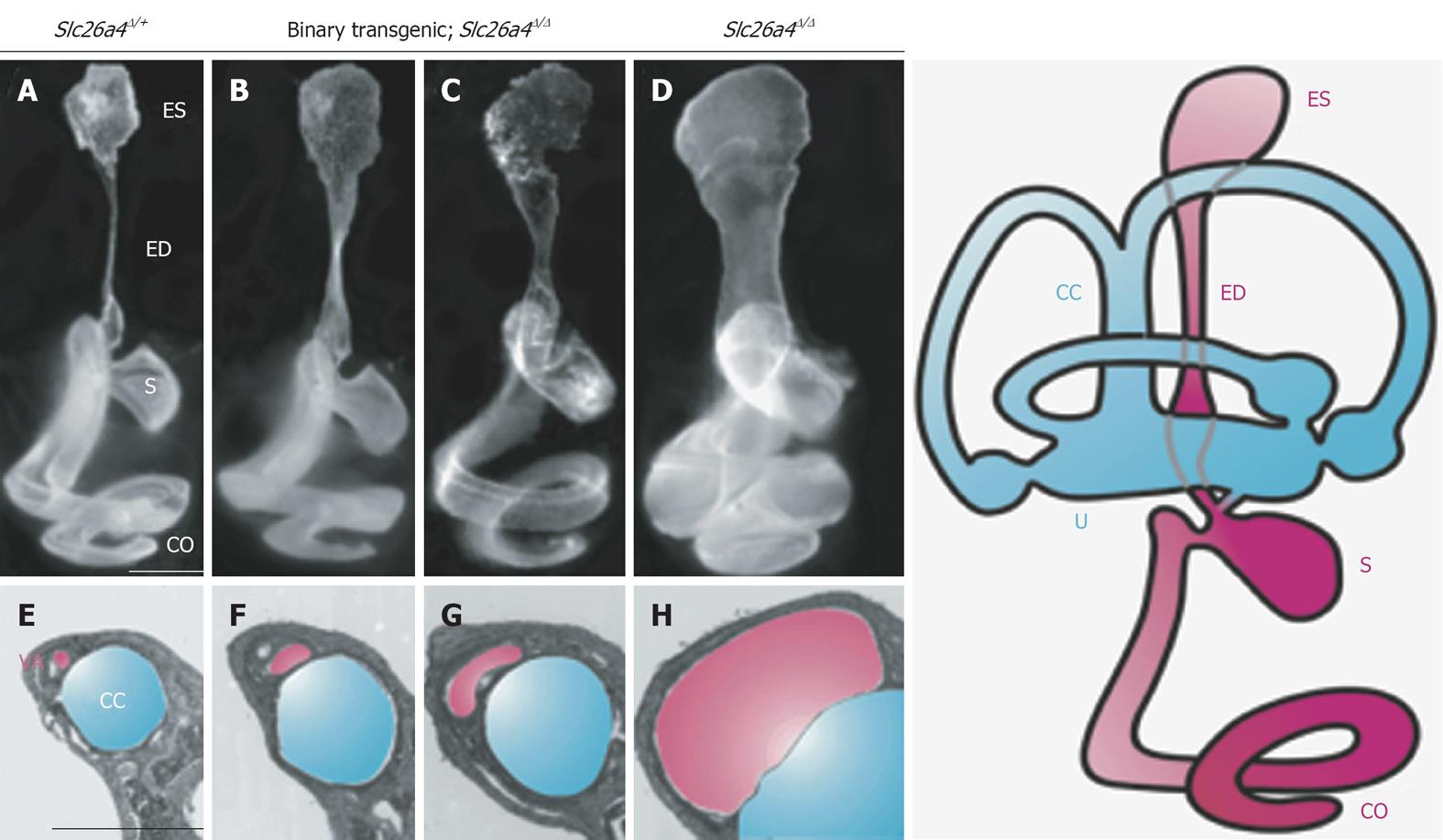
Figure 3 Morphology of the endolymphatic sac and duct and vestibular aqueduct in Slc26a4 mutant mouse models of enlargement of the vestibular aqueduct.
Slc26a4Δ/+ normal control (A and E), binary transgenic;Slc26a4Δ/Δ (B, C, F and G), or Slc26a4Δ/Δ mutant control mice (D and H) were sacrificed at P3 for paint-fill analysis (A-D) or between P28 and P109 for cross-sectional histopathology of the vestibular aqueduct (VA, shaded pink) adjacent to the common crus (CC, shaded blue; E–H). Scale bars: 500 μm (A, applies to A–D; E, applies to E–H). Manipulating pendrin expression in binary transgenic; Slc26a4Δ/Δ mice results in less enlargement of the endolymphatic duct and sac and vestibular aqueduct (B, C, F and G). ES: Endolymphatic sac; ED: Endolymphatic duct; S: Saccule; U: Utricle; CO: Cochlea. Reproduced with modification from Choi et al[24].
-
Citation: Ito T, Muskett J, Chattaraj P, Choi BY, Lee KY, Zalewski CK, King KA, Li X, Wangemann P, Shawker T, Brewer CC, Alper SL, Griffith AJ.
SLC26A4 mutation testing for hearing loss associated with enlargement of the vestibular aqueduct. World J Otorhinolaryngol 2013; 3(2): 26-34 - URL: https://www.wjgnet.com/2218-6247/full/v3/i2/26.htm
- DOI: https://dx.doi.org/10.5319/wjo.v3.i2.26


