Copyright
©The Author(s) 2015.
World J Ophthalmol. Feb 12, 2015; 5(1): 1-15
Published online Feb 12, 2015. doi: 10.5318/wjo.v5.i1.1
Published online Feb 12, 2015. doi: 10.5318/wjo.v5.i1.1
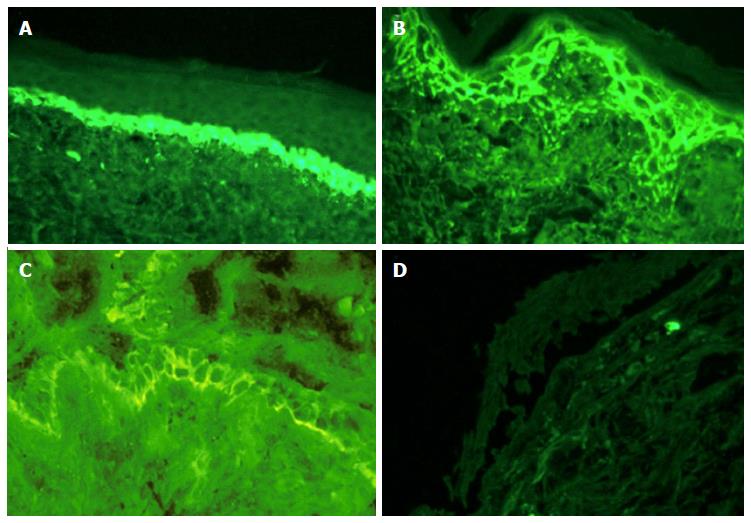
Figure 2 Direct immunofluorescence studies of conjunctival biopsies.
A: Conjunctival mucous membrane pemphigoid showing thick linear IgG along the lamina propria in a background of squamous metaplasia; B: Conjunctival pemphigus vulgaris showing linear IgG deposition on desmosomal areas of epithelial cell surfaces displaying a classic “chicken-wire” pattern; C: Conjunctival paraneoplastic pemphigus (PNP) showing linear IgG along the lamina propria with a hemidesmosomal pemphigoid-like in conjunction with a desmosomal pemphigus vulgaris-type epithelial cell surface “chicken-wire” type pattern. The pattern in PNP is due to the presence of IgG autoantibodies against hemidesmosomal antigens (plakin proteins: BP230/BPAG1 and plectin) as well as desmosomal antigens (plakin proteins: desmoplakin, envoplakin, periplakin, and desmogleins 3 and 1); D: Conjunctival pseudopemphigoid (most likely drug-induced) showing negative IgG deposition along the lamina propria in a background of subepithelial clefting, mild submucosal fibrosis, and incipient epithelial metaplasia.
- Citation: Huang LC, Wong JR, Alonso-Llamazares J, Nousari CH, Perez VL, Amescua G, Karp CL, Galor A. Pseudopemphigoid as caused by topical drugs and pemphigus disease. World J Ophthalmol 2015; 5(1): 1-15
- URL: https://www.wjgnet.com/2218-6239/full/v5/i1/1.htm
- DOI: https://dx.doi.org/10.5318/wjo.v5.i1.1


