Copyright
©The Author(s) 2017.
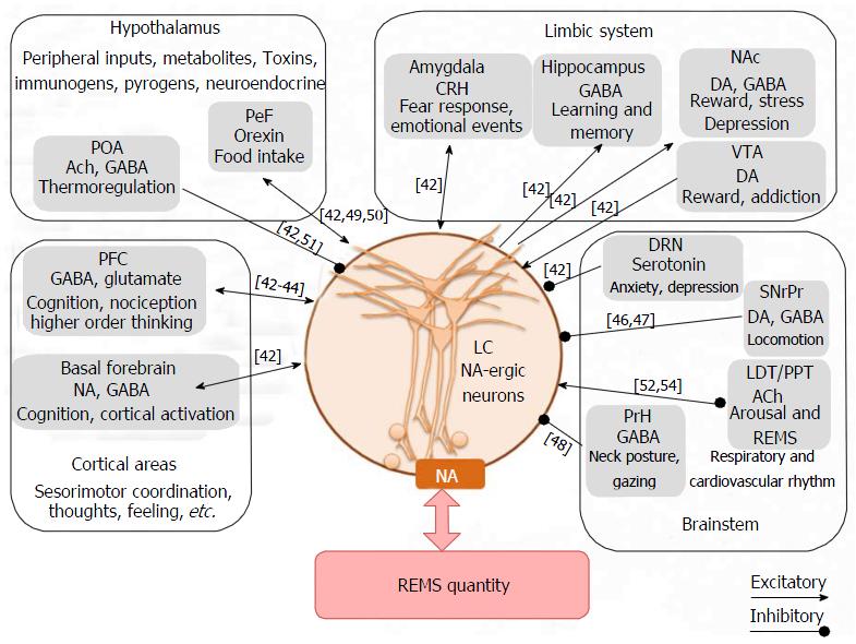
Figure 1 Schematic illustration of inputs to the locus coeruleus neurons from different regions in the brain, some of them are influenced by the environmental changes.
The resultant output of the LC neurons is responsible for level of NA in the brain. As those LC-NA-ergic neurons cease activity during REMS, disturbance in the latter keeps those neurons active and thus modulates NA level in the brain. This altered level of NA (mostly elevated level) in turn affects physiological processes regulated by the brain. PFC: Prefrontal cortex; NAc: Nucleus accumbens; VTA: Ventral tagmental area; DRN: Dorsal raphe nucleus; SNrPr: Substantia niagra pars reticulate; PrH: Prepositus hypoglossus; PeF: Perifornical area; POA: Preoptic area; LDT/PPT: Laterodorsal tegmentumpedunculopontine tegmentum; LC: Locus coeruleus; ACh: Acetylcholine; DA: Dopamine; GABA: Gamma-amino butyric acid; NA: Noradrenaline.
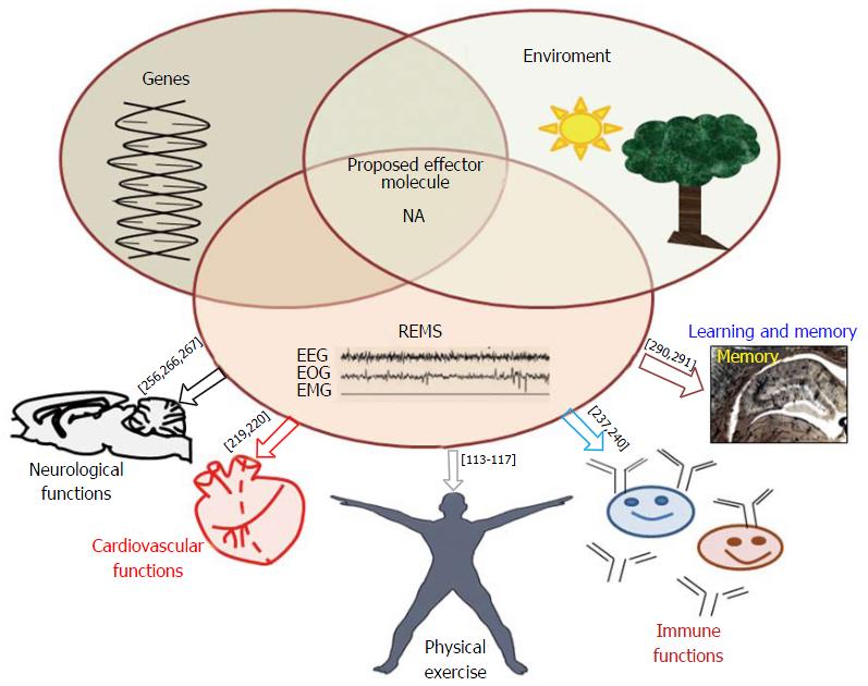
Figure 2 Interactions among environmental factors, rapid eye movement sleep and associated changes in gene expressions for long-term (sustained) modulation of patho-physiological changes have been diagrammatically represented in this figure.
NA is at least one of the common mediators to induce the changes, which ultimately affects the overall physiology in health and diseases. Abbreviations are as in the text. REMS: Rapid eye movement sleep; NA: Noradrenaline.
- Citation: Mehta R, Singh A, Mallick BN. Disciplined sleep for healthy living: Role of noradrenaline. World J Neurol 2017; 7(1): 6-23
- URL: https://www.wjgnet.com/2218-6212/full/v7/i1/6.htm
- DOI: https://dx.doi.org/10.5316/wjn.v7.i1.6


