Copyright
©2013 Baishideng Publishing Group Co.
Figure 1 Human brain from a middle-aged male, left hemisphere.
A: External surface; B: Internal surface. S: Supramarginal gyrus (Brodmann area 40); A: Angular gyrus (Brodmann area 39); F: Area of colors recognition (Brodmann area 19); N: Area of names recognition (Brodmann area 37); H: Area of auditory attention (effort to listen) (Brodmann area 21); M: Area of place memory (Brodmann area 19); V1: Cortex of the superior wall of the calcarine sulcus (Brodmann area 17); V2: Cortex of the inferior wall of the calcarine sulcus (Brodmann area 17).
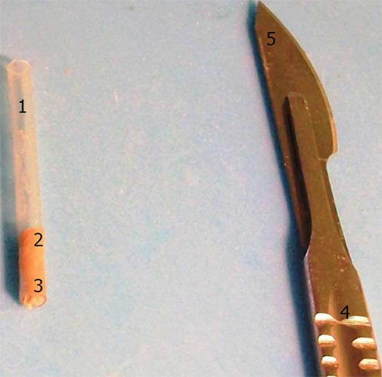
Figure 2 Method of cortical thickness measuring.
1: Plastic tube with brain sample; 2: Cerebral cortex within the tube; 3: Subcortical white matter within the tube; 4: Scalpel; 5: Scalpel blade.
- Citation: Mavridis I, Lontos K, Anagnostopoulou S. Thickness-based correlations of cortical areas involved in senses, speech and cognitive processes. World J Neurol 2013; 3(3): 67-74
- URL: https://www.wjgnet.com/2218-6212/full/v3/i3/67.htm
- DOI: https://dx.doi.org/10.5316/wjn.v3.i3.67


