
















Copyright: ©Author(s) 2026.
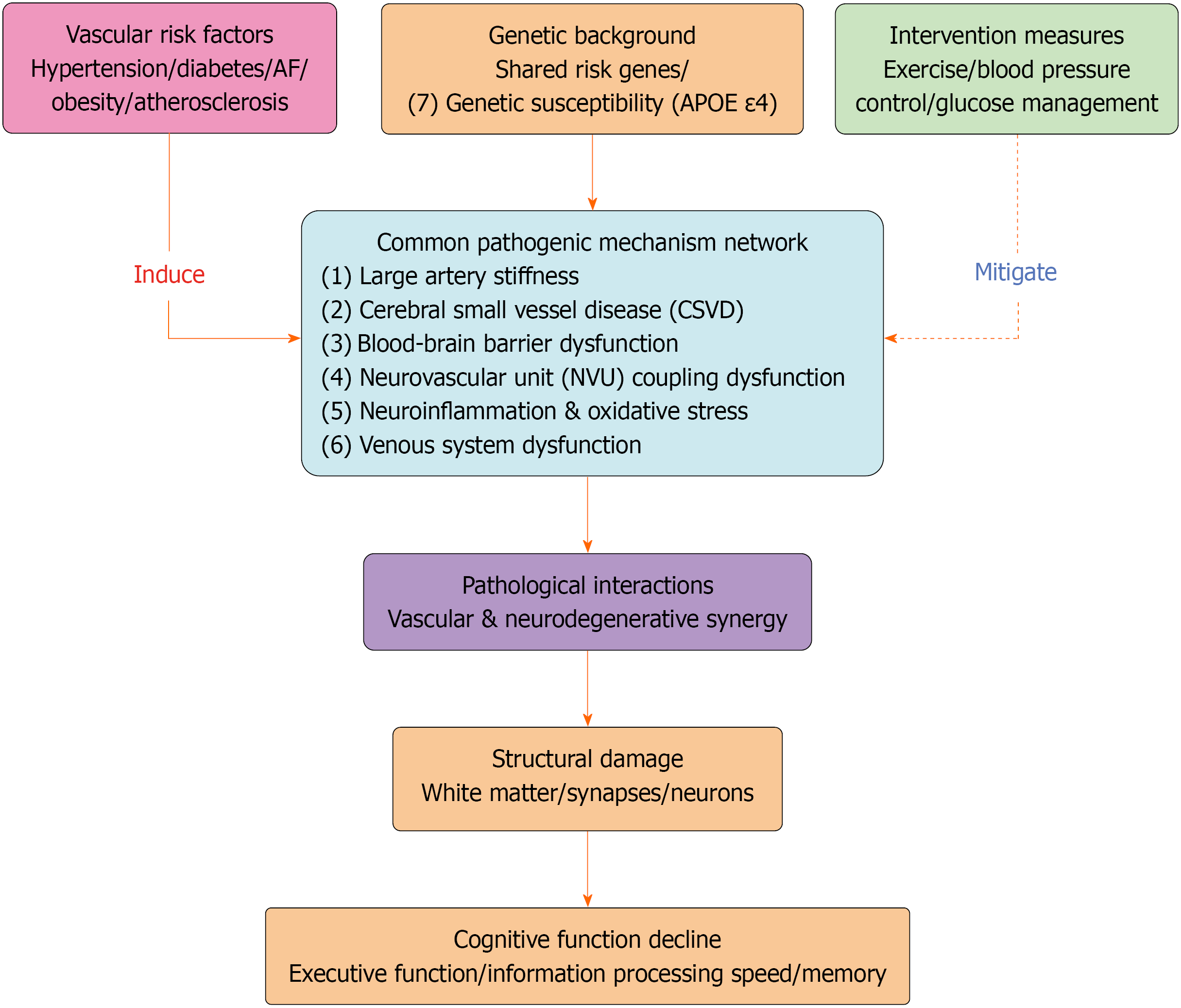
Figure 1 Integrated Pathomechanistic Network of Vascular Contributions to Cognitive Decline.
This figure illustrates the multi-layered pathogenic cascade through which vascular risk factors lead to cognitive impairment. Numbered pathways [(1)-(7)] represent key mechanistic nodes: (1) Large artery stiffness: Hypertension and atherosclerosis increase arterial stiffness, elevating systolic blood pressure and pulse pressure, which impairs cerebral perfusion and transmits damaging pulsatile stress to cerebral microvessels[55,56]; (2) Cerebral small vessel disease (CSVD): Vascular risk factors cause lipohyalinosis and fibrinoid necrosis of small arteries, leading to white matter hyperintensities, lacunar infarcts, cerebral microbleeds, and enlarged perivascular spaces, disrupting white matter tract integrity[39,41,57,72]; (3) Blood-brain barrier function and glymphatic impairment: Endothelial tight junction disruption increases permeability, allowing toxic blood components into brain parenchyma and impairing glymphatic clearance of neurotoxic proteins like β-amyloid[26-28,73]; (4) Neurovascular unit coupling failure: Endothelial dysfunction impairs neurovascular coupling, reducing cerebral blood flow response to neural activity and creating energy supply-demand mismatch[74,75,79]; (5) Neuroinflammation and oxidative stress: Microglial activation releases pro-inflammatory cytokines and generates reactive oxygen species, causing cellular damage and creating a self-perpetuating feedback loop that amplifies all mechanisms[58,76,77,80]; (6) Venous system dysfunction (emerging): Internal jugular vein reflux increases cerebral venous pressure, promoting vasogenic edema and Blood-brain barrier leakage, contributing to white matter damage[24,25]; and (7) Genetic susceptibility: Genetic factors, particularly apolipoprotein E epsilon 4 carriage, modulate vulnerability across all pathways by disrupting lipid homeostasis, promoting neuroinflammation, and exacerbating atherosclerosis[69]. These interconnected mechanisms converge to produce characteristic cognitive deficits including slowed processing speed, executive dysfunction, and memory impairment, culminating in vascular cognitive impairment, mixed dementia (Alzheimer’s disease + CSVD), or post-stroke cognitive impairment[5,7,35-38]. Furthermore, CSVD-related pathology contributes to neuropsychiatric symptoms such as depression and apathy, expanding the clinical impact of vascular brain injury[42]. AF: Atrial fibrillation; APOE ε4: Apolipoprotein E epsilon 4; CSVD: Cerebral small vessel disease; NVU: Neurovascular unit.
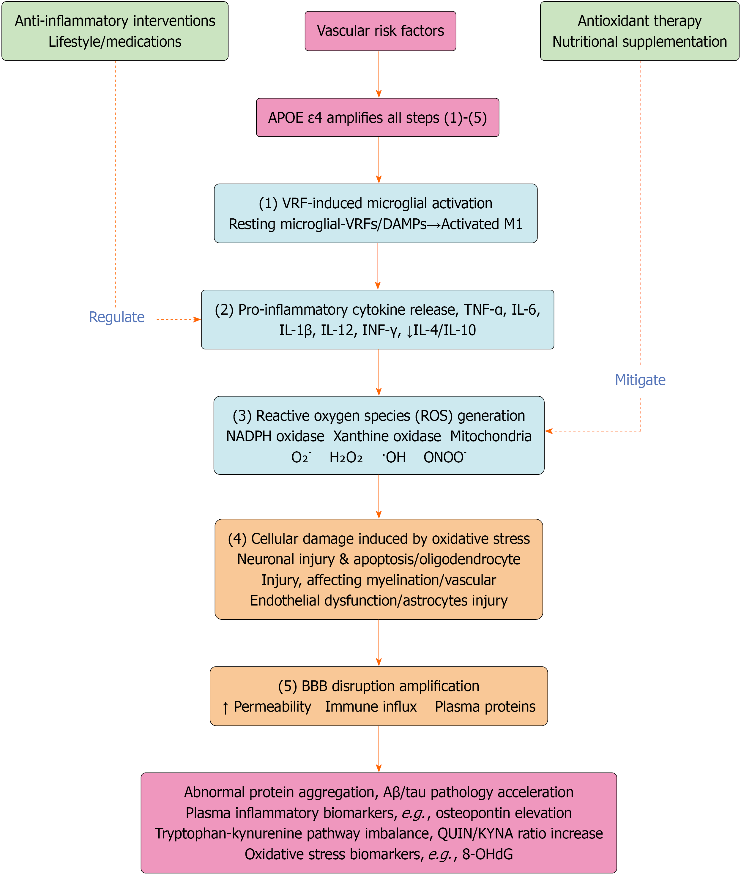
Figure 2 Mechanism Diagram of Neuroinflammation and Oxidative Stress.
This figure details the self-perpetuating cycle of neuroinflammation and oxidative stress that amplifies vascular injury. Numbered pathways [(1)-(5)] represent sequential and interconnected steps: (1) Microglial activation: Vascular risk factors and damage-associated molecular patterns trigger the transformation of resting ramified microglia into activated amoeboid M1 phenotype cells. Activation involves NOD-like receptor protein 3 inflammasome assembly, major histocompatibility complex class II upregulation, and increased phagocytic activity[58,76]; (2) Pro-inflammatory cytokine release: Activated microglia release a cascade of pro-inflammatory cytokines including tumor necrosis factor-alpha [endothelial activation, blood-brain barrier (BBB) disruption, neuronal apoptosis], interleukin (IL)-6 (acute phase response), IL-1β (microglial amplification, synaptic impairment), IL-12, and interferon-gamma while anti-inflammatory cytokines (IL-4, IL-10) are suppressed[76,77,80]; (3) Reactive oxygen species (ROS) generation: Cytokines trigger ROS production through multiple enzymatic sources (nicotinamide adenine dinucleotide phosphate oxidase, xanthine oxidase, uncoupled endothelial nitric oxide synthase, myeloperoxidase, inducible nitric oxide synthase) and non-enzymatic sources (mitochondrial electron transport chain, endoplasmic reticulum stress). ROS species (O2•−, H2O2, •OH, ONOO-) cause lipid peroxidation, protein oxidation, and DNA damage. Mitochondrial dysfunction creates a vicious cycle of further ROS production[58,76]; (4) Cellular damage: ROS and cytokines cause direct damage to multiple cell types: Neurons: Excitotoxicity, synaptic loss, apoptosis, tau hyperphosphory
- Citation: Jiang B. Vascular risk factors and cognitive decline: An integrated perspective from epidemiology to precision intervention. World J Neurol 2026; 12(1): 119653
- URL: https://www.wjgnet.com/2218-6212/full/v12/i1/119653.htm
- DOI: https://dx.doi.org/10.5316/wjn.119653