Copyright
©2013 Baishideng.
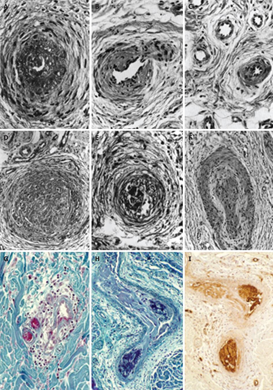
Figure 1 The histopathological appearances of a skin punch biopsy from areas of recently relapsed erythromelalgia 1 wk after aspirin discontinuation arteriolar l fibromuscular intimal proliferation, swollen endothelial cells, slight intravascular and perivascular infiltration with inflammatory cells, some perivascular fibrosis and no occlusive arteriolar thrombi.
The histopathological appearances from relapsed erythromelalgia complicated by blue ischemic spots 3 wk after aspirin discontinuation show arterioles with pronounced fibromuscular intimal proliferation without occlusive trombi (A-C) and fibromuscular intimal proliferation with occlusive thrombotic arteriolar lesions (D-F) perivascular oedema, vascular and perivascular fibrosis and infiltration by inflammatory cells (thromboangiitis obliterans). Originated from Michiels et al[20]. Biopsy from red-bluish discolored erythromelalgic skin area showing arteriolarthrombi with a weak fibrin staining (H) as compared to a positive fibrin staining from a patient with primary antipholipid syndrome and recurrent arterial skin thromboses (G). The occlusive arteriolar thrombi revealed a strong staining for von Willebrand factor (I) indicating platelet rich thrombi. This documents that thrombi from skin areas of erythromelalgia complicated by acrocyanosis are indeed platelet-rich and fibrin-poor thrombi. Originated from van Genderen et al[24].
- Citation: Michiels JJ, Ten Kate FW, Koudstaal PJ, Van Genderen PJ. Aspirin responsive platelet thrombophilia in essential thrombocythemia and polycythemia vera. World J Hematol 2013; 2(2): 20-43
- URL: https://www.wjgnet.com/2218-6204/full/v2/i2/20.htm
- DOI: https://dx.doi.org/10.5315/wjh.v2.i2.20


