Copyright
©2014 Baishideng Publishing Group Inc.
World J Hematol. Nov 6, 2014; 3(4): 118-127
Published online Nov 6, 2014. doi: 10.5315/wjh.v3.i4.118
Published online Nov 6, 2014. doi: 10.5315/wjh.v3.i4.118
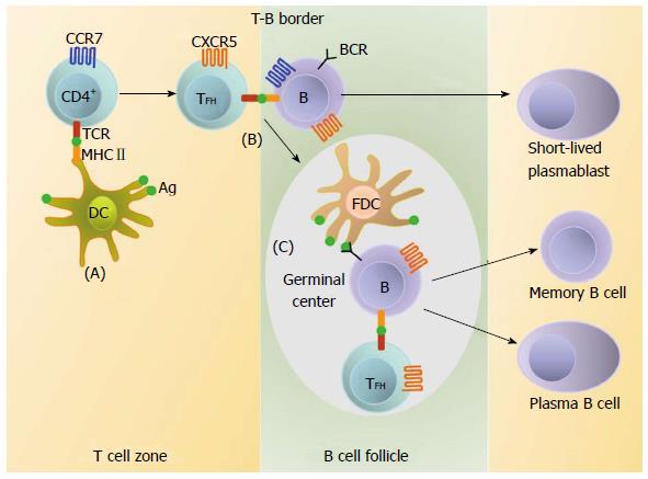
Figure 1 Follicular helper T cells and the differentiation program of B lymphocytes.
A: Naïve CD4+ T cells are activated following recognition of antigen (Ag) presented by dendritic cells (DC) in T cell zones. Upon antigen activation and co-stimulation by DC, nascent TFH upregulate CXC-chemokine receptor 5 (CXCR5), downregulate CC-chemokine receptor 7 (CCR7) and migrate towards B cell follicles; B: At the T-B border TFH contact antigen-activated B cells that move to the T-cell zone after upregulating CCR7. TFH deliver help to B cells resulting in their differentiation into short-lived extrafollicular plasmablasts or their migration into B cell-follicles to form germinal centers (GCs); C: Within GC, TFH promotes the B cell differentiation into long-lived plasma cells and memory B cells. TFH: Follicular helper T lymphocytes; FDC: Follicle dendritic cell; BCR: B cell receptor; MHC: Major histocompatibility complex; TCR: T cell receptor.
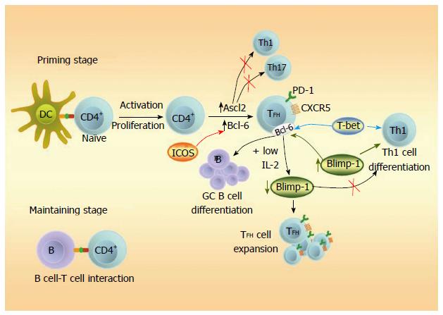
Figure 2 Regulation of Follicular helper T cell development.
After antigen-presenting signaling of dendritic cells (DC) to CD4+ T cells during priming, achaete-scute homologue 2 (Ascl2) and B cell lymphoma 6 (Bcl-6) induced by the inducible costimulator (ICOS), trigger for TFH differentiation program and inhibit Th1 and Th17 genes. Bcl-6 also controls germinal center (GC) B cell differentiation. B lymphocyte-induced maturation protein 1 (Blimp-1) and the T-box transcription factor (T-bet) regulate the function of Bcl-6 and favor the induction of a Th1 profile. Under low interleukin 2 (IL-2) conditions, excess of Bcl-6 counteracts Blimp-1 allowing expansion of the TFH and reduction of the Th1 programs of differentiation. Initial priming is sufficient to acquire the TFH markers but cognate B cells are needed for the subsequent maintenance stage. TFH: Follicular helper T lymphocytes; CXCR5: Chemokine (C-X-C motif) receptor 5; Th: T helper.
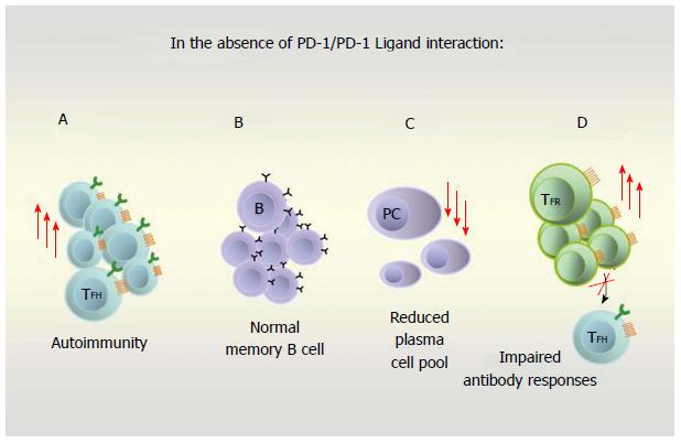
Figure 3 Inhibitory receptor programmed cell death 1, its ligands and their role in humoral response.
In the absence of an operative programmed cell death 1 (PD-1)/PD-1 Ligand axis, follicular helper T lymphocytes (TFH) increase and autoimmunity may develop (A); memory B cells are formed normally (B); reduced plasma cells (PC) are found (C); Foxp3+ CXCR5+ Bcl-6+ regulatory T cells (TFR) increase and have higher suppressive ability on TFH function leading to impaired antibody responses (D). PD-1: Programmed cell death-1; CXCR5: Chemokine (C-X-C motif) receptor; Bcl: B cell lymphoma.
- Citation: Parodi C, Badano MN, Galassi N, Coraglia A, Baré P, Malbrán A, Bracco MME. Follicular helper T lymphocytes in health and disease. World J Hematol 2014; 3(4): 118-127
- URL: https://www.wjgnet.com/2218-6204/full/v3/i4/118.htm
- DOI: https://dx.doi.org/10.5315/wjh.v3.i4.118


