Copyright
©2014 Baishideng Publishing Group Co.
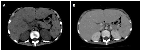
Figure 1 Computer tomography scan images of the liver.
Computed tomography (CT) scans of the liver performed on the 51st d (A) and 239th d (B) are presented.
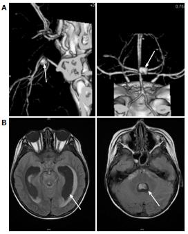
Figure 2 Three-dimensional reconstituted image of the intracranial aneurysm (A) and computer tomography scan (B).
A: Arrows indicate intracranial aneurysm; B: Arrows indicate dilated ventricles and subdural hemorrhage.
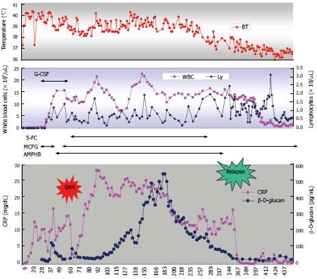
Figure 3 Clinical course of the patient.
Initiation of prednisolone was designated as day 1. WBC: White blood cell; 5-FC: 5-flucytosine; MCFG: Micafungin; CRP: C-reactive protein.
- Citation: Okawa T, Ono T, Endo A, Takagi M, Nagasawa M. Chronic disseminated candidiasis complicated with a ruptured intracranial fungal aneurysm in ALL. World J Hematol 2014; 3(2): 44-48
- URL: https://www.wjgnet.com/2218-6204/full/v3/i2/44.htm
- DOI: https://dx.doi.org/10.5315/wjh.v3.i2.44


