
















Copyright: ©Author(s) 2026.
World J Dermatol. Sep 16, 2026; 12(1): 117337
Published online Sep 16, 2026. doi: 10.5314/wjd.117337
Published online Sep 16, 2026. doi: 10.5314/wjd.117337
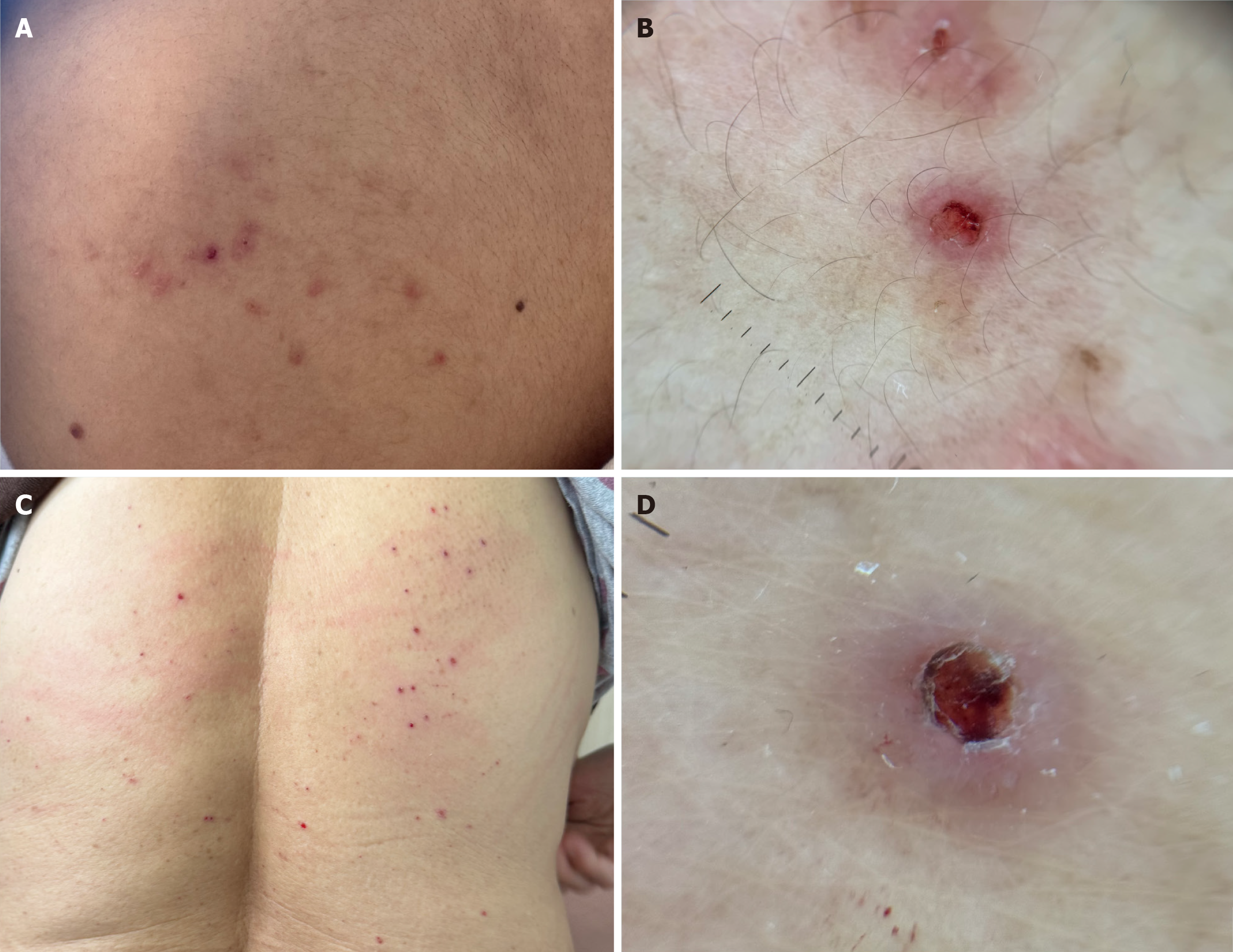
Figure 1 Clinicodermoscopic spectrum of pityriasis lichenoides.
A: Acute-spectrum presentation with multiple erythematous-brown papules, several showing central crusts at different evolutionary stages on the extremity; B: Dermoscopy of an acute-spectrum lesion showing a central reddish-brown amorphous/crusted area with a peripheral white collarette of scale and dotted/glomerular vessels; C: Chronic papular presentation pityriasis lichenoides chronica C-like (PLC-like) with fine scaling on the back; D: Dermoscopy of a PLC-like lesion showing a central brown-crusted plug over a yellow-brown structureless background with peripheral fine scale and sparse vessels. These findings align with recent dermoscopic descriptions and may aid recognition without biopsy.
- Citation: Kaya G. Clinicopathological spectrum of pityriasis lichenoides: A case series of 19 patients. World J Dermatol 2026; 12(1): 117337
- URL: https://www.wjgnet.com/2218-6190/full/v12/i1/117337.htm
- DOI: https://dx.doi.org/10.5314/wjd.117337