
















Copyright: ©Author(s) 2026.
World J Anesthesiol. Sep 22, 2026; 14(1): 123553
Published online Sep 22, 2026. doi: 10.5313/wja.123553
Published online Sep 22, 2026. doi: 10.5313/wja.123553
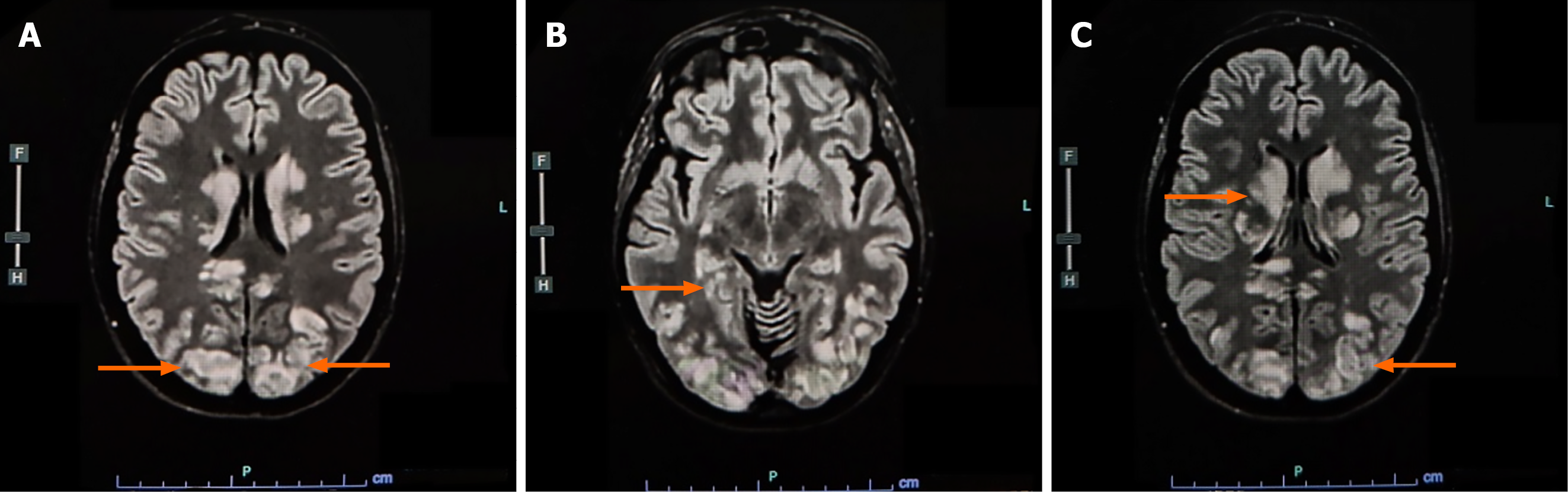
Figure 1 Axial fluid-attenuated inversion recovery magnetic resonance images.
A: Symmetric extensive bilateral cortico-subcortical hyperintensities involving the parieto-occipital regions within the posterior circulation territories; B: Extension of hyperintense lesions into deep central structures, including the basal ganglia and periventricular white matter; C: Diffuse bilateral vasogenic edema with widespread cortical-subcortical hyperintensities involving both posterior and central brain regions.
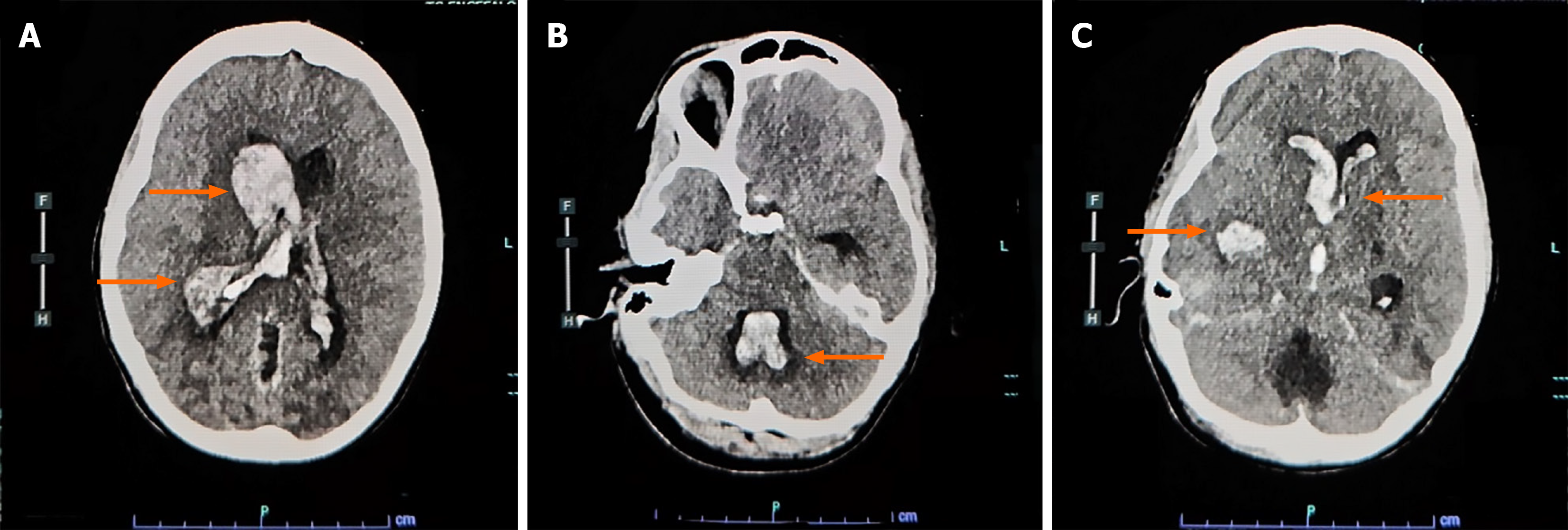
Figure 2 Cranial computed tomography images.
A: Massive intraventricular hemorrhage involving the lateral and third ventricles, associated with diffuse cerebral edema and ventricular enlargement; B: Blood extension into the fourth ventricle at the level of the posterior fossa, with marked brainstem compression and obliteration of the basal cisterns, findings consistent with severe intracranial hypertension; C: Right capsuloganglionic intraparenchymal hemorrhage with massive intraventricular extension, surrounding edema, hydrocephalus, and significant mass effect.
- Citation: Merciai F, Pagano T, Petrosino M, Erra A, Scarano D, Muoio R, Carbone D. Fulminant postpartum posterior reversible encephalopathy syndrome associated with hemolysis, elevated liver enzymes, and low platelet count syndrome: A case report. World J Anesthesiol 2026; 14(1): 123553
- URL: https://www.wjgnet.com/2218-6182/full/v14/i1/123553.htm
- DOI: https://dx.doi.org/10.5313/wja.123553