Copyright
©The Author(s) 2017.
World J Clin Oncol. Aug 10, 2017; 8(4): 360-365
Published online Aug 10, 2017. doi: 10.5306/wjco.v8.i4.360
Published online Aug 10, 2017. doi: 10.5306/wjco.v8.i4.360
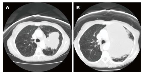
Figure 1 Computed tomography scan of the chest showing the left lung mass.
A: At the time of diagnosis; B: Explosive growth of the tumor after two cycles of chemotherapy.
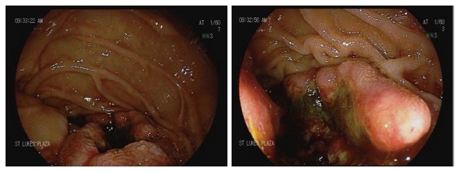
Figure 2 Esophagogastroduodenoscopy showing the malignant-appearing 1-cm mass in the second part of the duodenum.
The scope could not traverse the lesion and the exam could not be finished. Cold forceps biopsies were taken for histology.
- Citation: Qasrawi A, Tolentino A, Abu Ghanimeh M, Abughanimeh O, Albadarin S. BRAF V600Q-mutated lung adenocarcinoma with duodenal metastasis and extreme leukocytosis. World J Clin Oncol 2017; 8(4): 360-365
- URL: https://www.wjgnet.com/2218-4333/full/v8/i4/360.htm
- DOI: https://dx.doi.org/10.5306/wjco.v8.i4.360


