Copyright
©The Author(s) 2016.
World J Clin Oncol. Feb 10, 2016; 7(1): 114-121
Published online Feb 10, 2016. doi: 10.5306/wjco.v7.i1.114
Published online Feb 10, 2016. doi: 10.5306/wjco.v7.i1.114
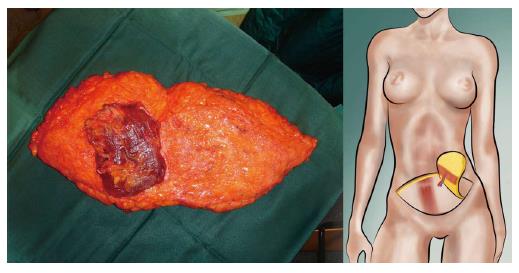
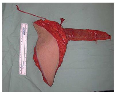
Figure 1 Typical example of a free transverse rectus abdominis muscle-flap showing the undersurface with its vascular pedicle and the completely excised rectus abdominis muscle.
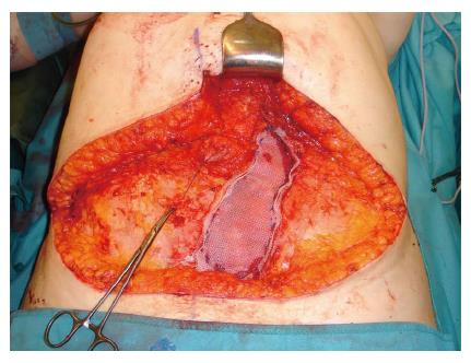
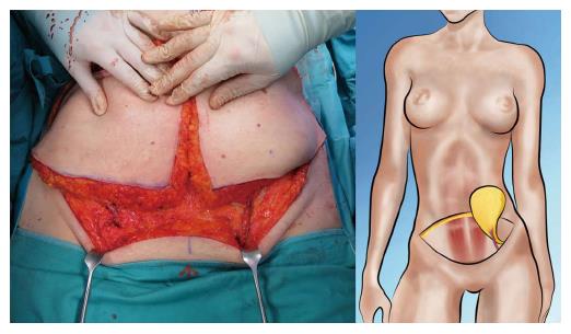
Figure 2 After harvest of a full transverse rectus abdominis muscle-flap, the fascial defect of the rectus fascia usually has to be closed with a synthetic mesh.
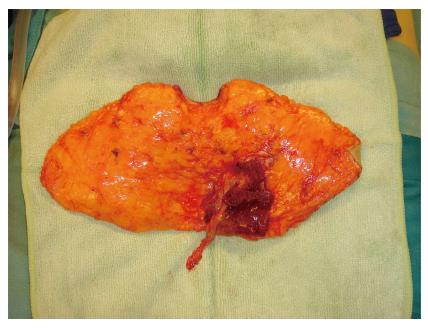
Figure 3 In a muscle-sparing free transverse rectus abdominis muscle, only a portion of the rectus muscle is included in the flap.
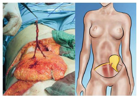
Figure 4 Example of a deep inferior epigastric perforator-flap with 2 perforators merging into the deep inferior epigastric vessels.
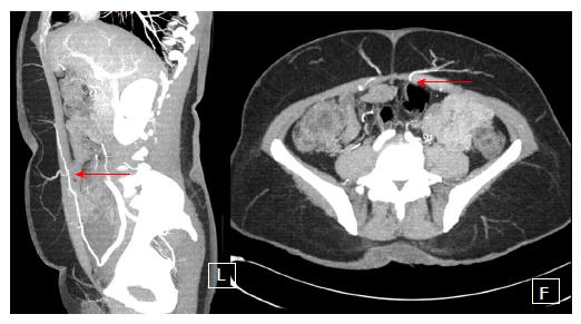
Figure 5 Computed tomographic-angiography to better elucidate the exact anatomy of perforators of the deep inferior epigastric perforator-vessels.
The red arrow points out the piercing of the perforator through the rectus abdominis muscle on the sagittal (left) and transverse (right) view.
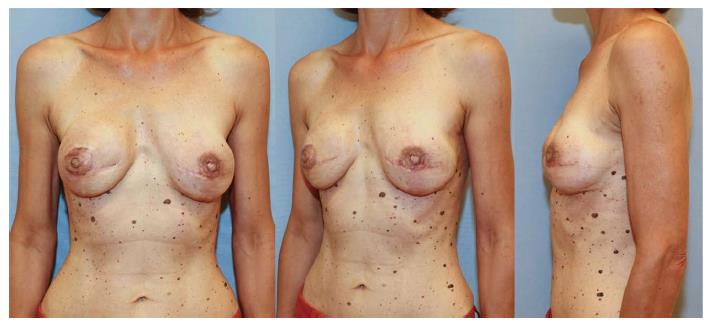
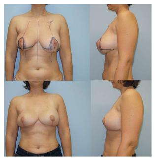
Figure 6 Pre- and post-operative pictures of a patient who underwent delayed reconstruction on her right side with a hemi-deep inferior epigastric perforator-flap and immediate reconstruction of her left side with the second hemi-deep inferior epigastric perforator-flap.
She already had reconstruction of the nipple-areola-complex with tattooing and a local skin flap.
Figure 7 Example of a split superficial inferior epigastric artery-flap with both pedicles clearly visible.
Since there is no injury to the rectus fascia, this the most desirable choice of flap from the lower abdomen.
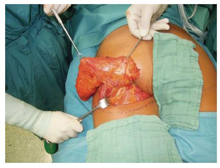
Figure 8 Example of a fasciocutaneous infragluteal-flap almost completely raised.
At the undersurface of the flap, the flap’s main vessels, i.e., the descending branch of the inferior gluteal vessels, can be seen clearly. Additionally, a branch of the posterior femoral cutaneous nerve is visible and spared.
Figure 9 A 49-year-old patient after bilateral nipple-sparing prophylactic mastectomy and immediate breast reconstruction with bilateral fasciocutaneous infragluteal-flap.
Figure 10 Intraoperative image of a completely harvested transverse myocutaneous gracilis flap with flap pedicle (short) and the relatively long saphenous vein as a venous supercharge.
Figure 11 Pre- and post-operative pictures after immediate bilateral skin-reducing breast reconstruction with a transverse musculocutaneous gracilis-flap and reconstruction of the nipple-areola-complex.
- Citation: Pollhammer MS, Duscher D, Schmidt M, Huemer GM. Recent advances in microvascular autologous breast reconstruction after ablative tumor surgery. World J Clin Oncol 2016; 7(1): 114-121
- URL: https://www.wjgnet.com/2218-4333/full/v7/i1/114.htm
- DOI: https://dx.doi.org/10.5306/wjco.v7.i1.114


