Copyright
©The Author(s) 2025.
World J Clin Oncol. Aug 24, 2025; 16(8): 108371
Published online Aug 24, 2025. doi: 10.5306/wjco.v16.i8.108371
Published online Aug 24, 2025. doi: 10.5306/wjco.v16.i8.108371
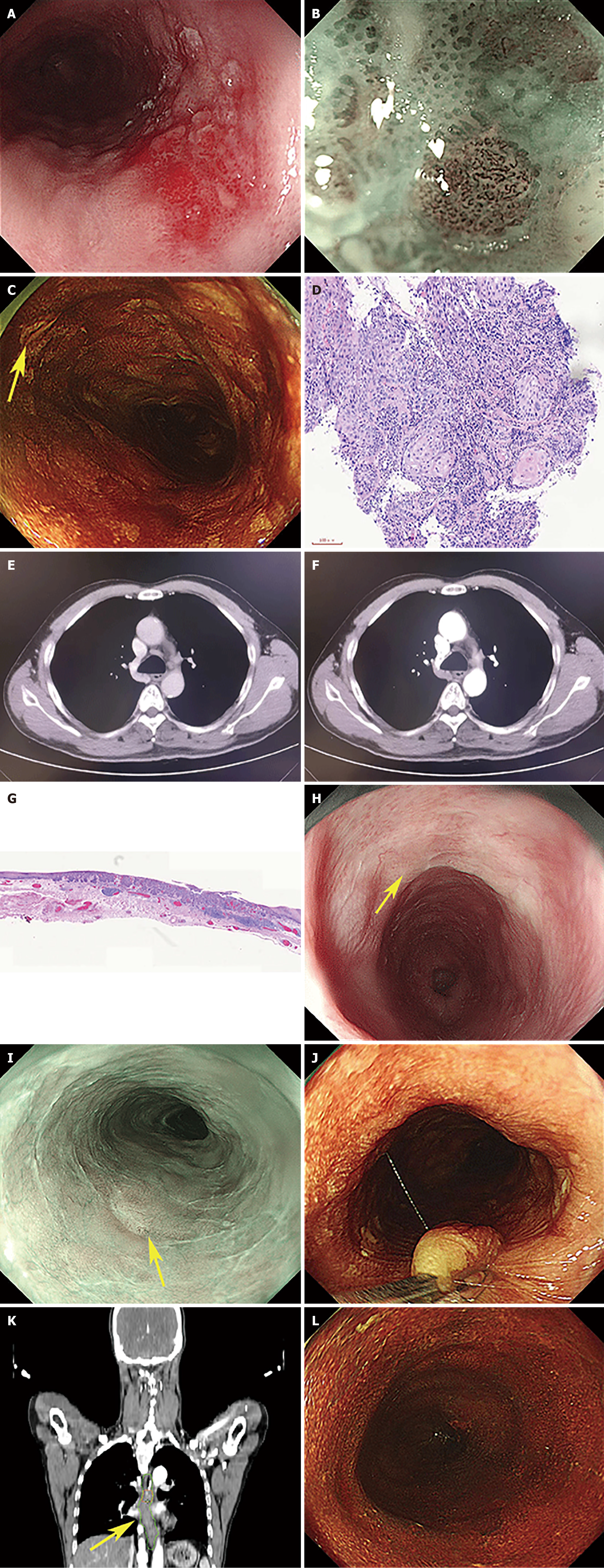
Figure 1 Imaging examinations.
A: Gastroscopy showing a slightly elevated reddish area; B: Narrow band imaging (NBI) magnifying gastroscopy findings; C: More than 10 Lesions were found per endoscopic view and defined as grade C; D: Biopsy pathology findings; E: Computed tomography in the venous phase; F: Computed tomography in the arterial phase; G: Postoperative pathological results; H: Endoscopic submucosal dissection (ESD) scar under the white light; I: ESD scar under NBI; J: Titanium clips were used for accurate marking on oral and anal sides for multiple Lugol-voiding lesions (LVL) ranges; K: Clinical target volume; L: Multiple LVL disappeared.
- Citation: Chen D, Zhong DF, Liu D. Exploration of preventive treatment for high risk patients with metachronous multiple esophageal squamous cell carcinoma: A case report. World J Clin Oncol 2025; 16(8): 108371
- URL: https://www.wjgnet.com/2218-4333/full/v16/i8/108371.htm
- DOI: https://dx.doi.org/10.5306/wjco.v16.i8.108371


