Copyright
©The Author(s) 2025.
World J Clin Oncol. Jul 24, 2025; 16(7): 107246
Published online Jul 24, 2025. doi: 10.5306/wjco.v16.i7.107246
Published online Jul 24, 2025. doi: 10.5306/wjco.v16.i7.107246
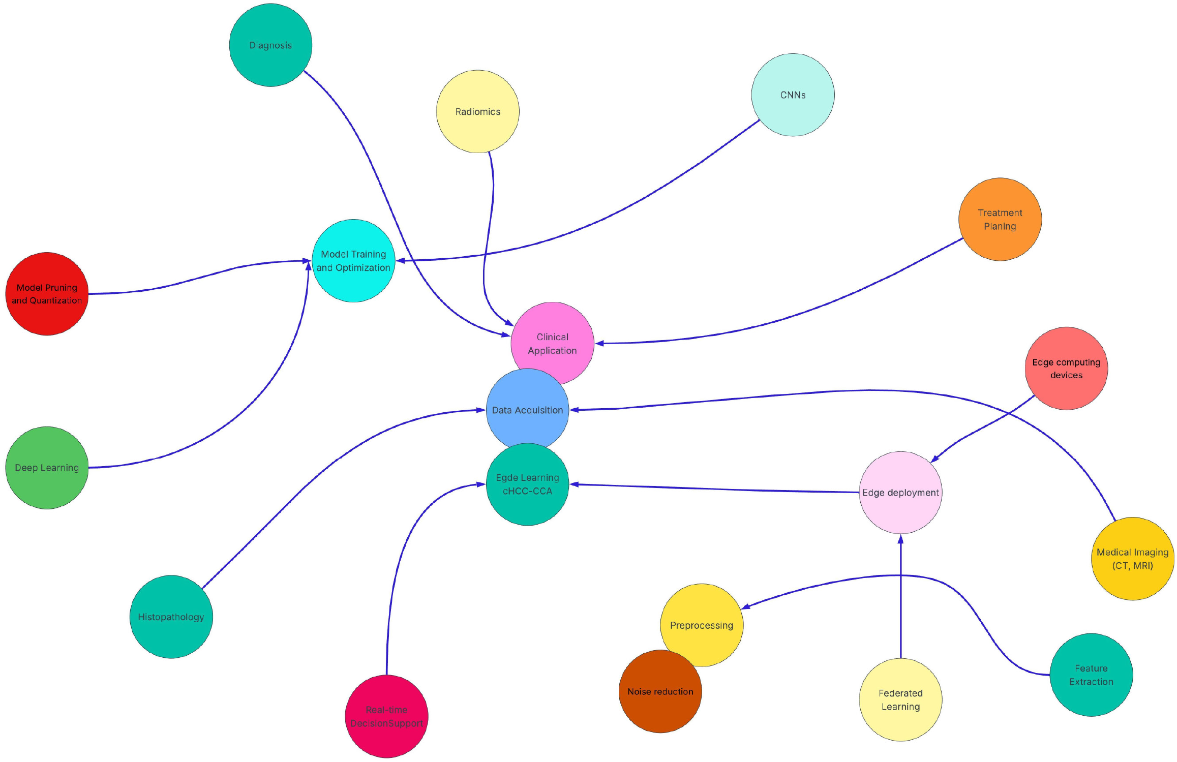
Figure 1 This diagram outlines the edge learning workflow for diagnosing and classifying combined hepatocellular-cholangiocarcinoma.
The process begins with data acquisition (computed tomography, magnetic resonance imaging, histology, radiomics), followed by preprocessing (noise reduction, feature extraction) to enhance data quality. Deep learning models, including convolutional neural networks, pruning, and quantization, optimize efficiency and accuracy. Trained models are deployed on edge devices using federated learning, ensuring real-time inference and data privacy. Clinically, edge learning enhances diagnosis, treatment planning, and intraoperative decision support, improving patient outcomes. Color-coded nodes represent key stages, with blue arrows indicating process flow. Edge learning boosts efficiency, safeguards privacy, and enables real-time decision-making, transforming combined hepatocellular-cholangiocarcinoma management. CT: Computed tomography; MRI: Magnetic resonance imaging; CNNs: Convolutional neural networks; cHCC-CCA: Combined hepatocellular-cholangiocarcinoma.
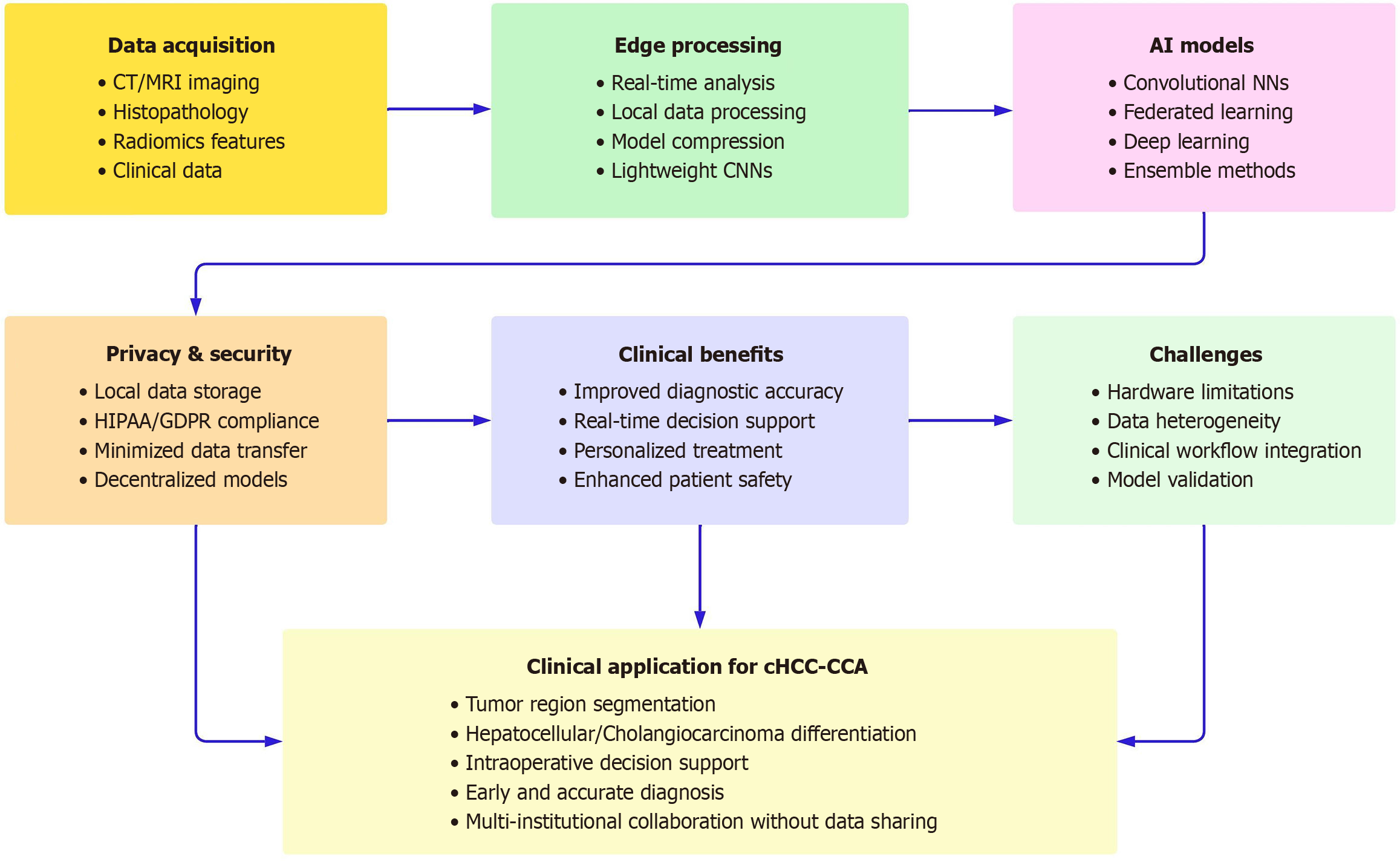
Figure 2 The key aspects of edge learning applications in combined hepatocellular-cholangiocarcinoma diagnosis and classification.
CT: Computed tomography; MRI: Magnetic resonance imaging; CNNs: Convolutional neural networks; AI: Artificial intelligence; HIPAA: Health insurance portability and accountability act; GDPR: General data protection regulation.
- Citation: Akbulut S, Colak C. Edge learning applications in the prediction and classification of combined hepatocellular-cholangiocarcinoma: A comprehensive narrative review. World J Clin Oncol 2025; 16(7): 107246
- URL: https://www.wjgnet.com/2218-4333/full/v16/i7/107246.htm
- DOI: https://dx.doi.org/10.5306/wjco.v16.i7.107246


