Copyright
©The Author(s) 2025.
World J Clin Oncol. May 24, 2025; 16(5): 105444
Published online May 24, 2025. doi: 10.5306/wjco.v16.i5.105444
Published online May 24, 2025. doi: 10.5306/wjco.v16.i5.105444
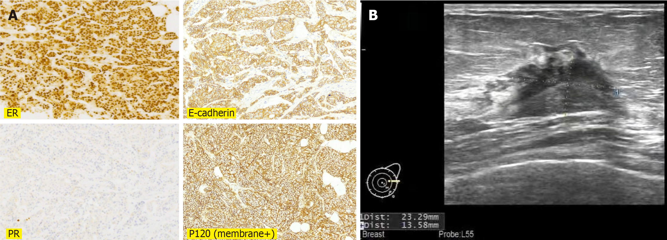
Figure 1 Invasive ductal carcinoma of the breast.
A: Immunohistochemistry revealed ER (90%), E-cadherin (+), PR (5%), P120 (membrane+), indicating a diagnosis of invasive ductal carcinoma of the breast; B: Breast ultrasound showed a solid mass in the left breast with a diameter of 2.4 cm.
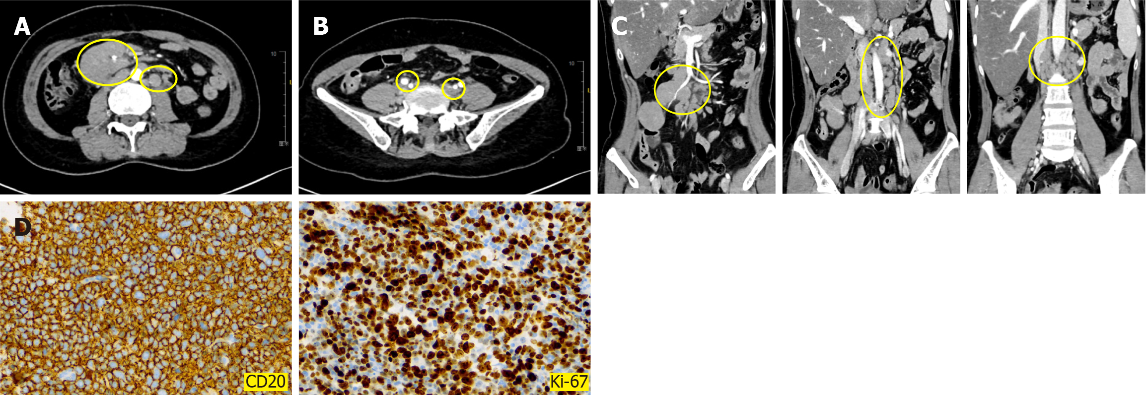
Figure 2 Metachronous diffuse large B-cell lymphoma.
A-C: Computed tomography showed multiple enlarged lymph nodes adjacent to the abdominal aorta (A) and bilateral iliac vessels (B) and abdominal cavity (C); D: Immunohistochemical results showed that CD20 (+), Ki-67 (80%). These results suggested a diagnosis of diffuse large B-cell lymphoma.
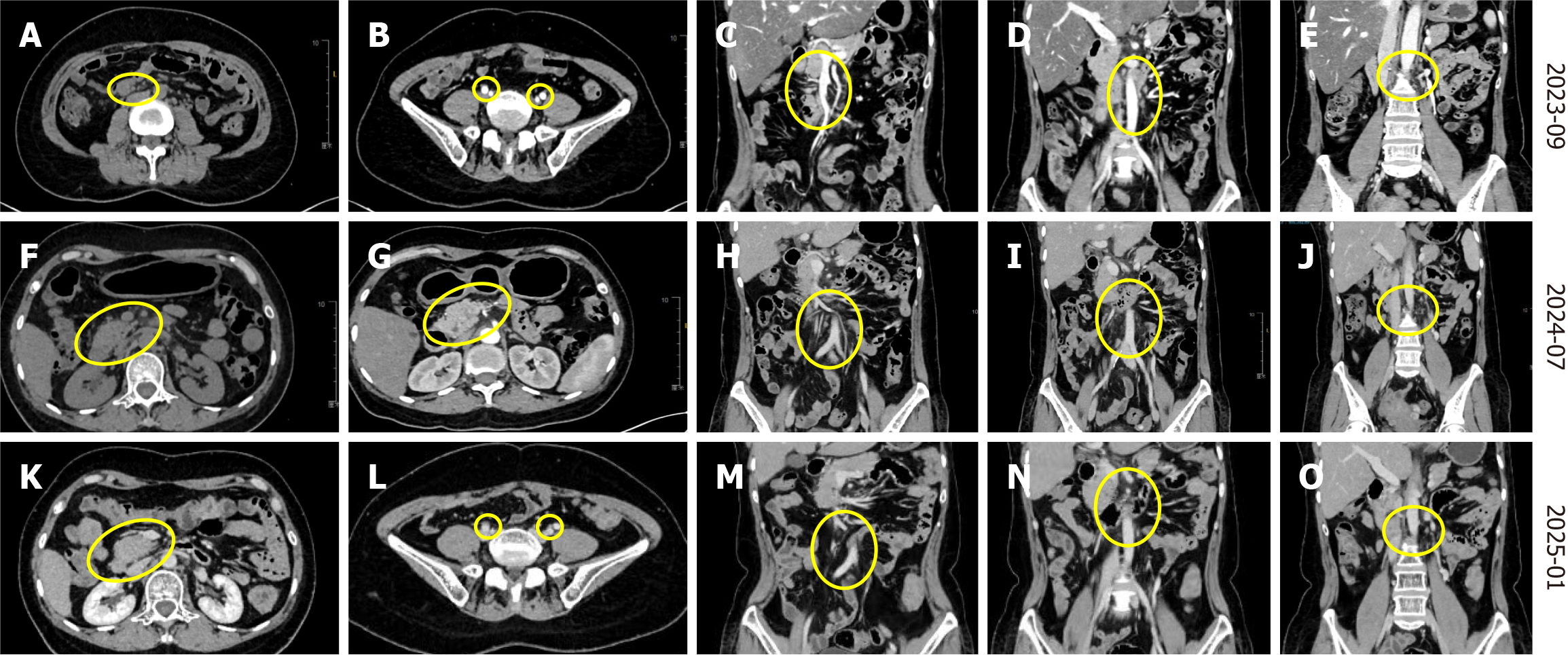
Figure 3 Treatment outcome of diffuse large B-cell lymphoma.
A-E: Lymph nodes adjacent to the abdominal aorta (A) and bilateral iliac vessels (B) and abdominal cavity (C-E) were significantly reduced compared to before treatment; F and G: Multiple lymph node shadows can be seen near the abdominal aorta, bilateral iliac vessels, and abdominal cavity, partially fused into clusters; H-J: The abdominal lymph nodes were significantly shrunk and decreased compared to before treatment; K-O: The latest re-examination result shows that lymph nodes adjacent to the abdominal aorta (K) and bilateral iliac vessels (L) and abdominal cavity (M-O) were significantly reduced.
- Citation: Luo M, Liu RN, He ZM, Liang QF, Huang FL. Diagnosis and treatment of metachronous multiple primary carcinoma: A case report and review of literature. World J Clin Oncol 2025; 16(5): 105444
- URL: https://www.wjgnet.com/2218-4333/full/v16/i5/105444.htm
- DOI: https://dx.doi.org/10.5306/wjco.v16.i5.105444


