Copyright
©The Author(s) 2025.
World J Clin Oncol. May 24, 2025; 16(5): 104398
Published online May 24, 2025. doi: 10.5306/wjco.v16.i5.104398
Published online May 24, 2025. doi: 10.5306/wjco.v16.i5.104398
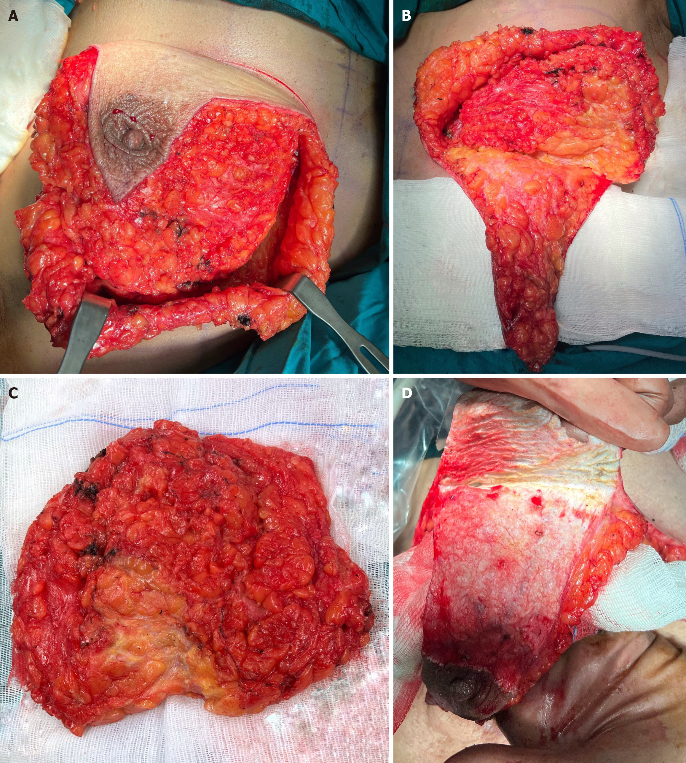
Figure 1 Surgical steps.
A: Skin incisions and breast tissue dissection; B: Mobilization of the flap with nipple-areolar complex; C: Breast tissue specimen; D: De-epithelization of the skin of the inferior mammary fold.
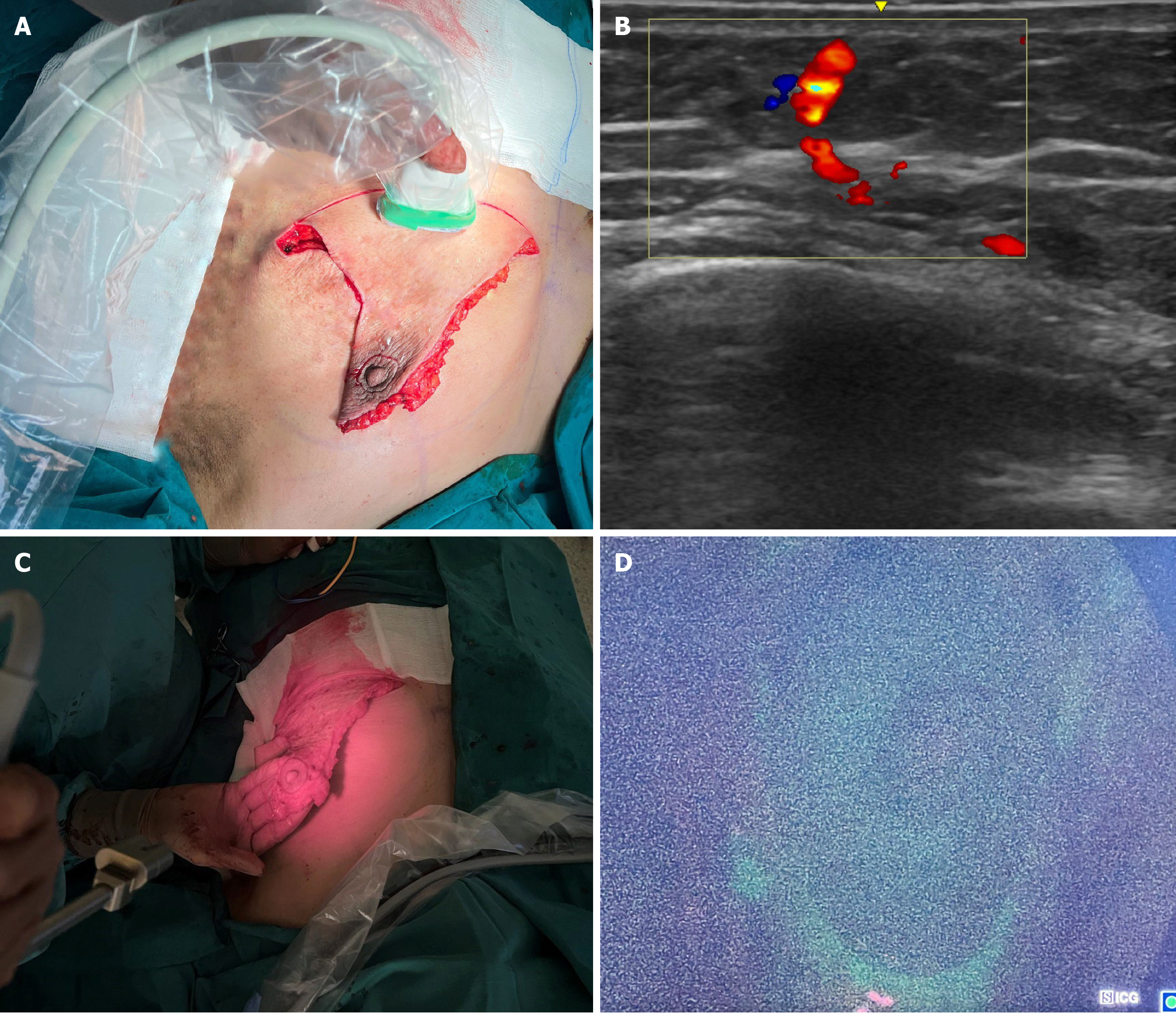
Figure 2 Auxiliary methods for tissue perfusion assessment.
A: Intraoperative Doppler ultrasonography (USG); B: USG image; C: Intraoperative fluorescence angiography with indocyanine green; D: Fluorescence imaging of the lower de-epithelized flap.
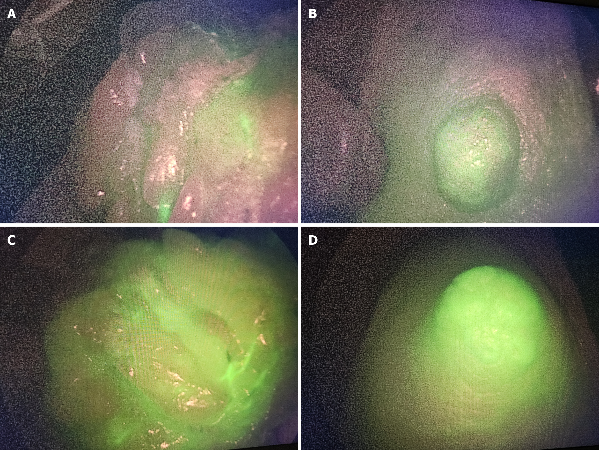
Figure 3 Assessment of blood supply with indocyanine green.
A: Subcutaneous tissue under nipple-areolar complex (NAC) 1 minute after indocyanine green (ICG) administration; B: NAC 1 minute after ICG administration; C: Subcutaneous tissue under NAC 3 minutes after ICG administration; D: NAC 3 minutes after ICG administration.
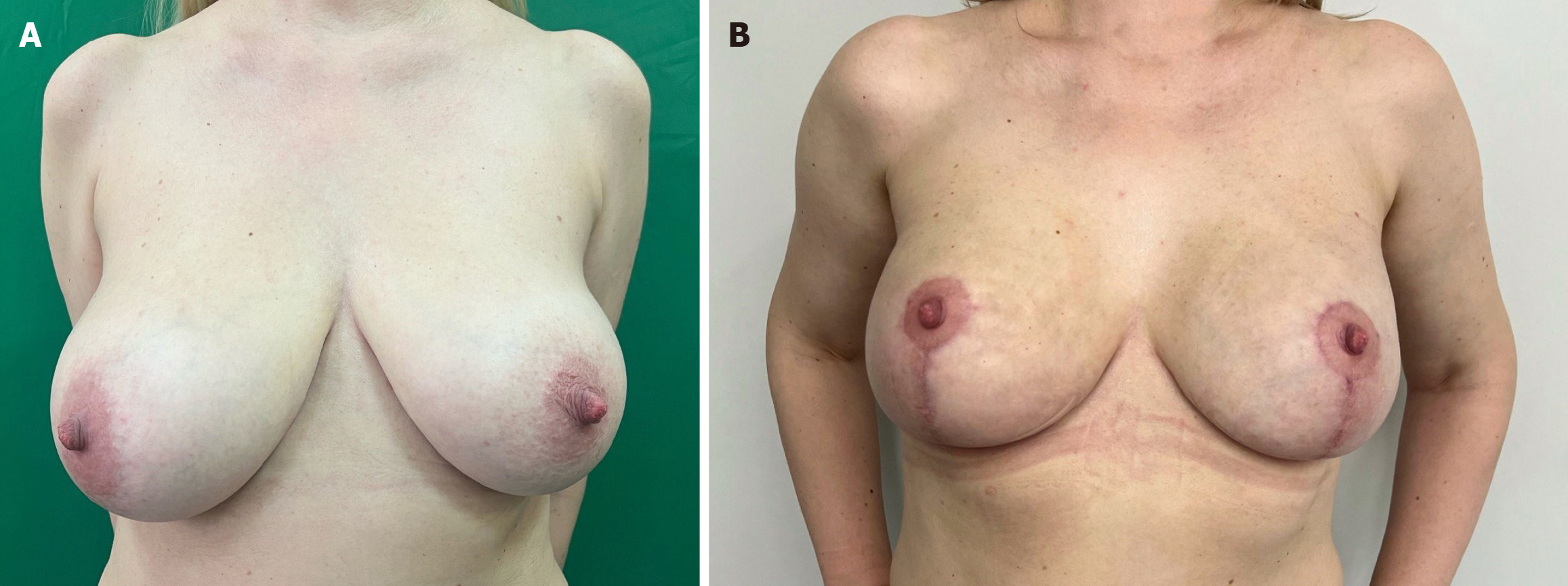
Figure 4 Preoperative and postoperative image.
A: Preoperative; B: 3 months postoperative.
- Citation: Sukhotko AS, Bumbu A, Covantsev S. Preservation of the nipple-areolar complex during subcutaneous mastectomy: A surgical and diagnostic method. World J Clin Oncol 2025; 16(5): 104398
- URL: https://www.wjgnet.com/2218-4333/full/v16/i5/104398.htm
- DOI: https://dx.doi.org/10.5306/wjco.v16.i5.104398


