Copyright
©The Author(s) 2024.
World J Clin Oncol. May 24, 2024; 15(5): 635-643
Published online May 24, 2024. doi: 10.5306/wjco.v15.i5.635
Published online May 24, 2024. doi: 10.5306/wjco.v15.i5.635
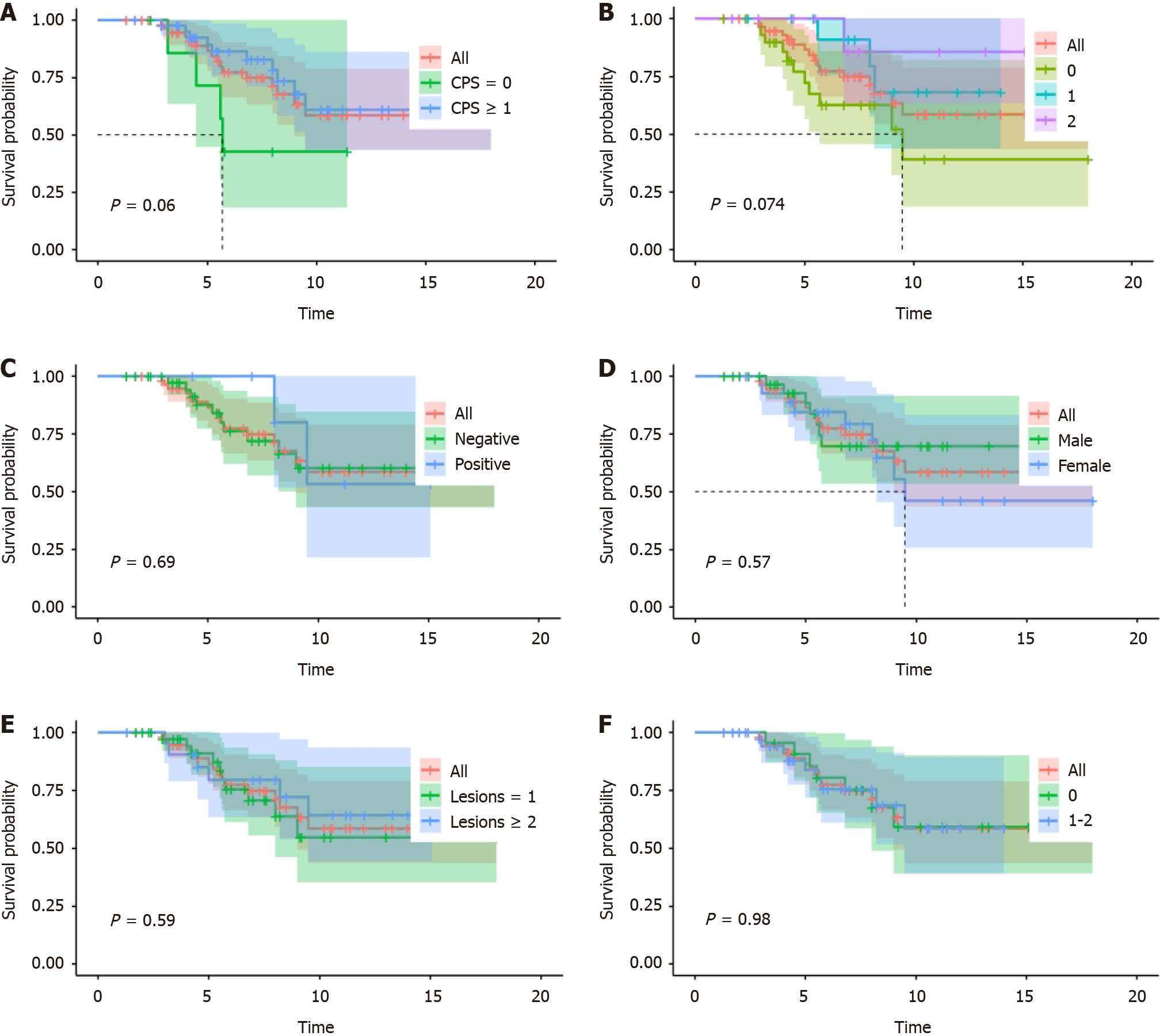
Figure 1 Progression-free survival of clinicopathological factors.
A: The progression-free survival (PFS) of different combined positive scores of programmed cell death 1 ligand 1; B: PFS according to different human epidermal growth factor receptor 2 expression levels; C: PFS according to Epstein-Barr virus-encoded small RNA status; D: PFS according to patient gender; E: PFS according to different numbers of metastatic organs; F: PFS according to different performance status scores.
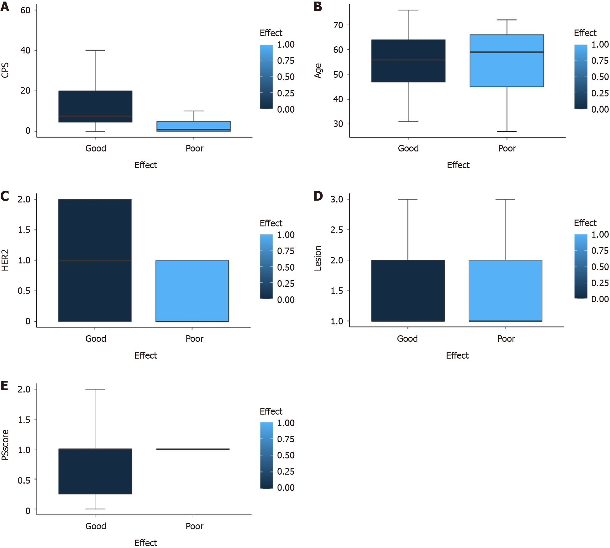
Figure 2 Risk factor analysis of clinicopathological factors.
A: Risk factor analysis of programmed cell death 1 Ligand 1 combined positive score scores; B: Risk factor analysis of patient age; C: Risk factor analysis of different numbers of metastatic organs; D: Risk factor analysis of human epidermal growth factor receptor 2 expression; E: Risk factor analysis of performance status scores. PS: Performance status; CPS: Combined positive score; HER2: Human epidermal growth factor receptor 2.
- Citation: Ma XT, Ou K, Yang WW, Cao BY, Yang L. Human epidermal growth factor receptor 2 expression level and combined positive score can evaluate efficacy of advanced gastric cancer. World J Clin Oncol 2024; 15(5): 635-643
- URL: https://www.wjgnet.com/2218-4333/full/v15/i5/635.htm
- DOI: https://dx.doi.org/10.5306/wjco.v15.i5.635


