Copyright
©The Author(s) 2024.
World J Clin Oncol. Mar 24, 2024; 15(3): 456-463
Published online Mar 24, 2024. doi: 10.5306/wjco.v15.i3.456
Published online Mar 24, 2024. doi: 10.5306/wjco.v15.i3.456
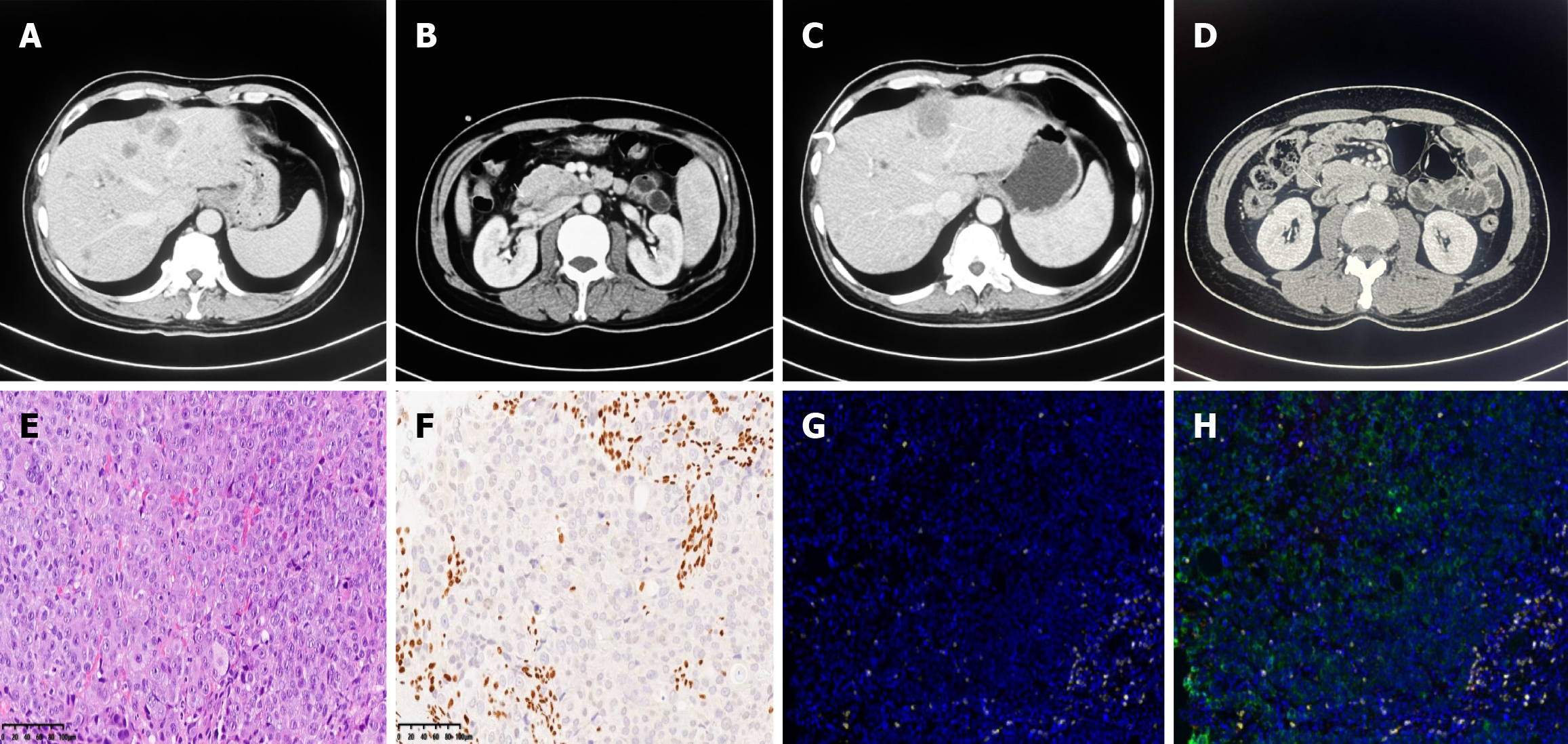
Figure 1 No response to immunochemotherapy or disease progression was confirmed by contrast computed tomography in case 1.
A and B: A mass in the descending part of the duodenum and multiple liver metastases at the end of 1st-line chemotherapy; C and D: Disease progression was confirmed by enlargement of liver metastases and infusions and impaired liver function with persisting mixed jaundice even after percutaneous transhepatic cholangial drainage; E: H&E: The undifferentiated tumor cells grew in cords, nests, and diffuse sheets, and the nucleus of the tumor was vacuolated with obvious nucleoli. Mitotic images and scattered multinucleated giant cells were also observed. Extensive areas of necrosis were observed within the tumor; F: The SMARCA4 protein was negative for BRG1 according to immunohistology; G: CD8+ T-cell infiltration was assessed by multiple immunofluorescence assays; H: Tertiary lymphoid structures were not intact, and case 1 was classified as an acquired immune tolerance type by microenvironment analysis.
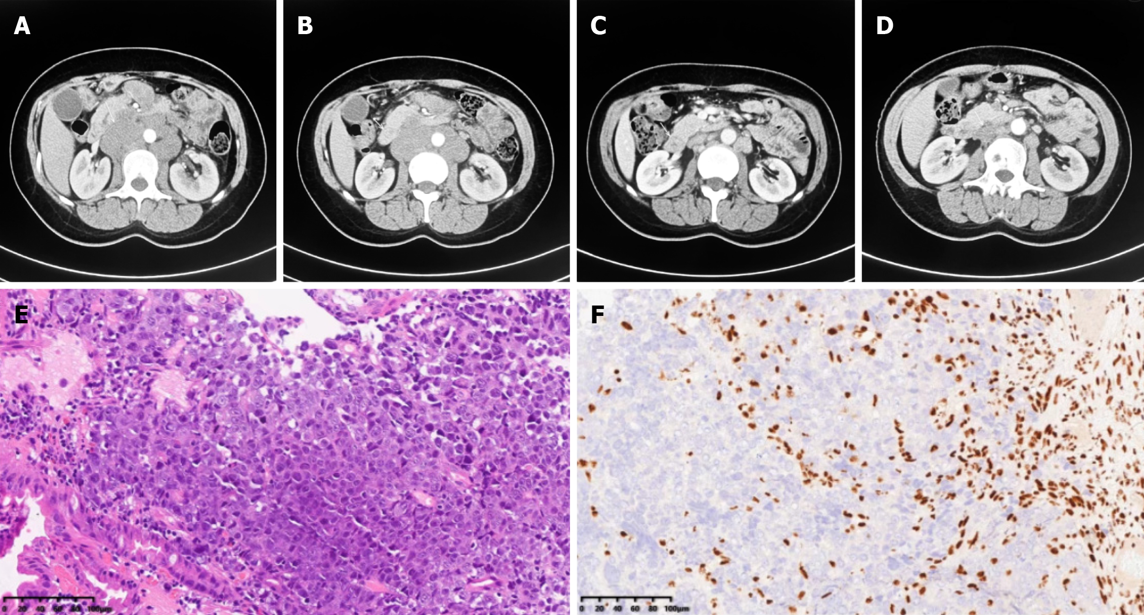
Figure 2 Major response to immunochemotherapy and disease regression in case 2.
A and B: A mass in the descending part of the duodenum and multiple retroperitoneal lymph node metastases initially observed; C and D: Significant disease regression after the administration of immunochemotherapy; E: H&E: The undifferentiated tumor cells were diffusely distributed, and patchy necrosis was observed; F: The SMARCA4 protein was negative for BRG1 according to immunohistology.
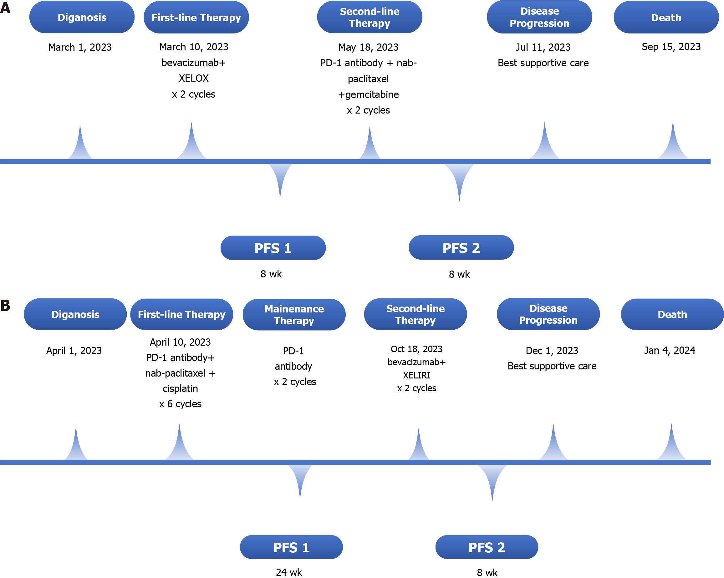
Figure 3 Timeline summarizing.
A: Case 1; B: Case 2. PFS: Progression-free survival; PFS1: PFS time during first-line therapy; PFS2: PFS time during second-line therapy.
- Citation: Shi YN, Zhang XR, Ma WY, Lian J, Liu YF, Li YF, Yang WH. PD-1 antibody in combination with chemotherapy for the treatment of SMARCA4-deficient advanced undifferentiated carcinoma of the duodenum: Two case reports. World J Clin Oncol 2024; 15(3): 456-463
- URL: https://www.wjgnet.com/2218-4333/full/v15/i3/456.htm
- DOI: https://dx.doi.org/10.5306/wjco.v15.i3.456


