Copyright
©The Author(s) 2023.
World J Clin Oncol. Apr 24, 2023; 14(4): 171-178
Published online Apr 24, 2023. doi: 10.5306/wjco.v14.i4.171
Published online Apr 24, 2023. doi: 10.5306/wjco.v14.i4.171
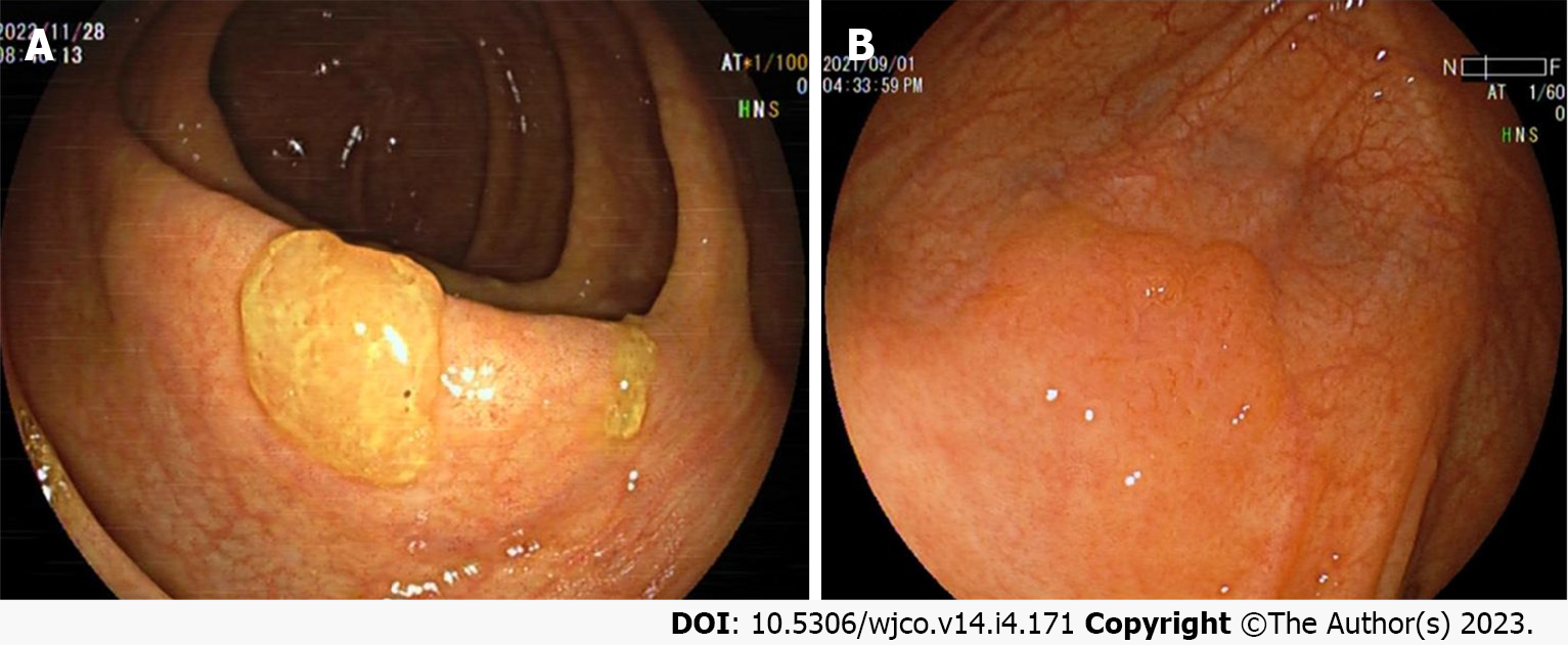
Figure 1 White light endoscopic features of colorectal sessile serrated lesion cases.
A: The sessile serrated lesions (SSLs) case with mucus cap under white light endoscopy; B: The borders of SSLs are not clearly distinguishable from the surrounding mucosa, and the morphology are cloud-like surface under white light. The above figure shows a case of SSL-D which has a reddish surface and a central depression.
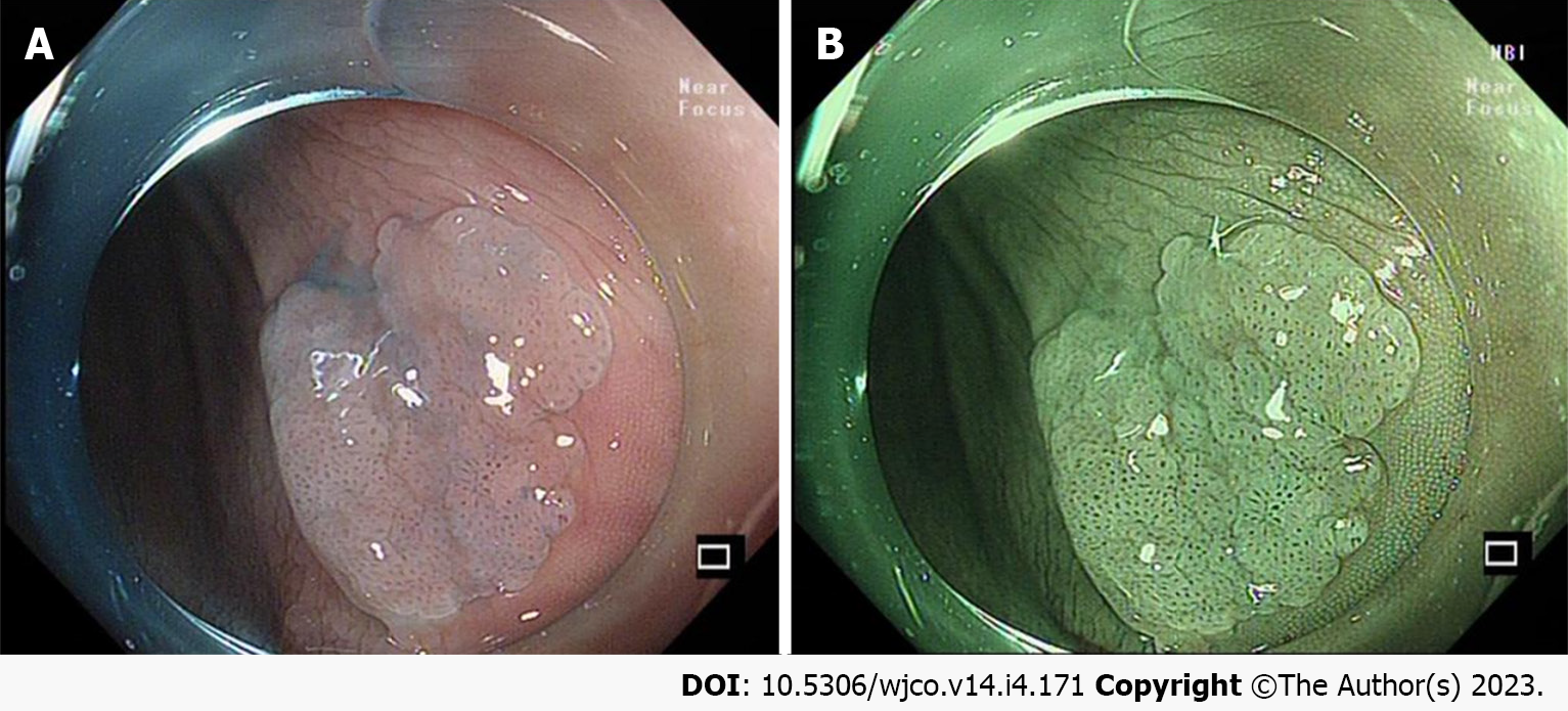
Figure 2 Endoscopic features of the colorectal sessile serrated lesion case after acetic acid spray.
A: The border of the sessile serrated lesions (SSLs) is clearly revealed under acetic acid spray; B: The combined application of acetic acid spray and narrow band imaging can clearly show the borders and surface microstructure of SSLs, which is more conducive to endoscopic treatment.
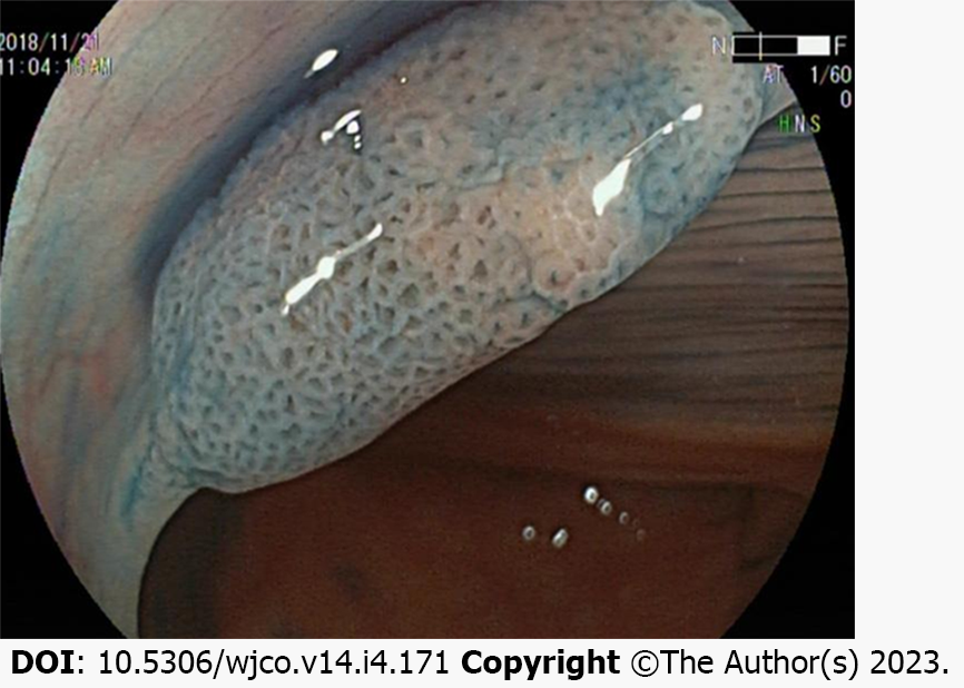
Figure 3 Endoscopic features of the colorectal sessile serrated lesion case after indigo carmine spray.
The opening pattern of the Pit II-O gland, features an expanded and more rounded shape, dilation of the colorectal sessile serrated lesion surface crypt.
Figure 4 Endoscopic features of colorectal sessile serrated lesion cases under narrow band imaging mode.
A: The mucus cap of sessile serrated lesions (SSLs) shows a brick-red appearance under narrow band imaging (NBI); B: The expansion of the surface crypt in the SSLs shows a black spot under NBI.
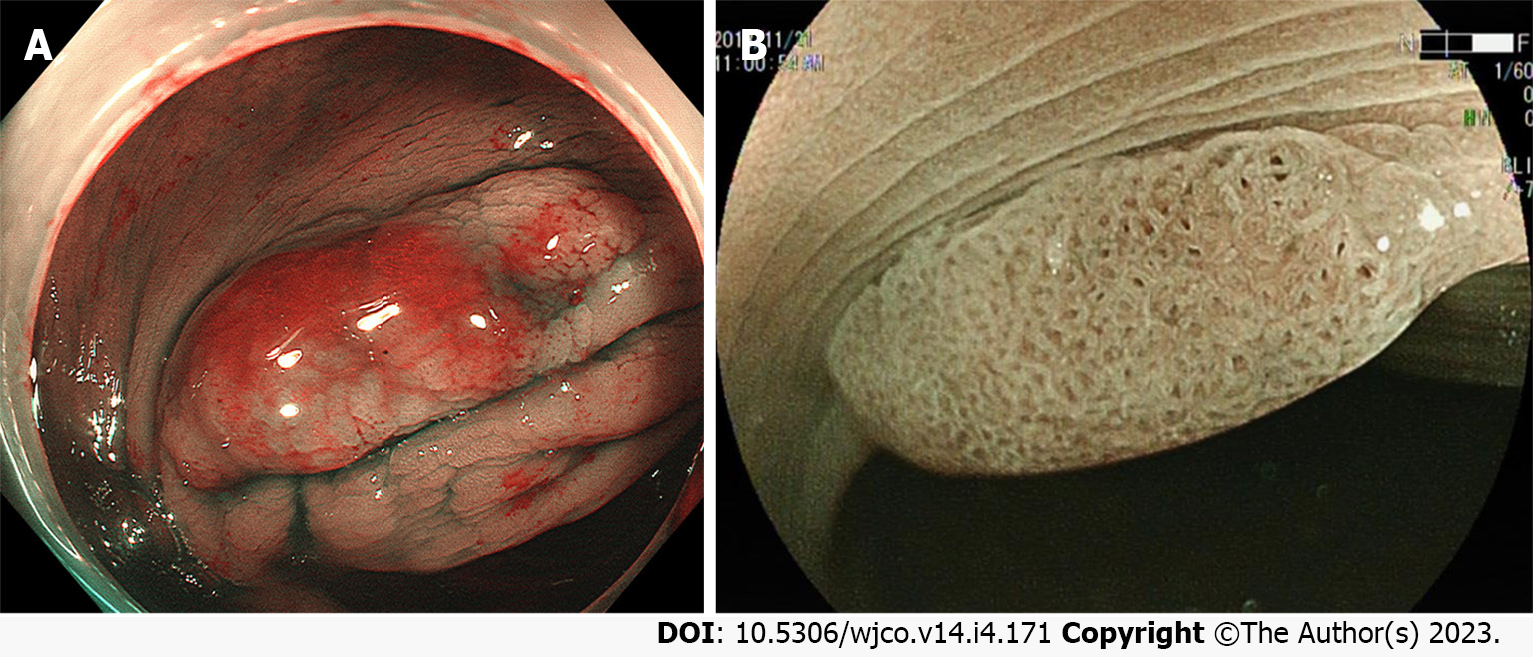
Figure 5 Endoscopic features of the colorectal sessile serrated lesion case under chromoscopy combined with magnified endoscopy.
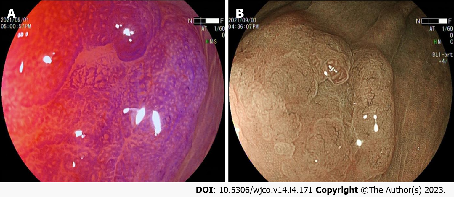
A: Crystalline violet spray makes the surface glandular structure of colorectal sessile serrated lesions (SSLs) more visible, and combined with magnified endoscopic observation is useful for inferring the pathological characteristics of the lesion; B: Blue light imaging combined with magnified endoscopic observation of the microstructure of the SSL surface revealed that the SSL-D case have a Pit III and/or Pit IV type of glandular duct opening pattern based on Pit II-O, and varicose microvessels on the surface of the lesion are found.
- Citation: Wang RG, Wei L, Jiang B. Current progress on the endoscopic features of colorectal sessile serrated lesions. World J Clin Oncol 2023; 14(4): 171-178
- URL: https://www.wjgnet.com/2218-4333/full/v14/i4/171.htm
- DOI: https://dx.doi.org/10.5306/wjco.v14.i4.171


