Copyright
©The Author(s) 2023.
World J Clin Oncol. Mar 24, 2023; 14(3): 131-137
Published online Mar 24, 2023. doi: 10.5306/wjco.v14.i3.131
Published online Mar 24, 2023. doi: 10.5306/wjco.v14.i3.131
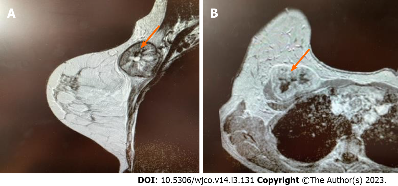
Figure 1 Breast magnetic resonance imaging.
Heterogeneous lesion 47 mm (orange arrows) on the border of the upper quadrants of the right breast with central zone of necrosis and peripheral vascularization. A: Axial; B: Frontal.
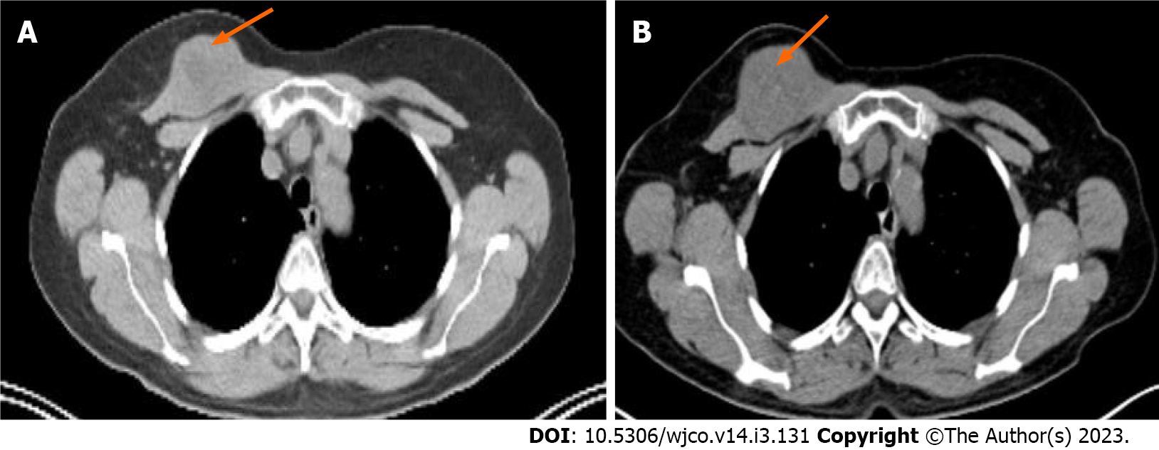
Figure 2 Positron emission tomography-computed tomography.
A: Computed tomography (CT) on September 2020; B: СТ on February 2021 demonstrated progression in the right breast lesion (orange arrows), size increased from 39 mm to 48 mm.
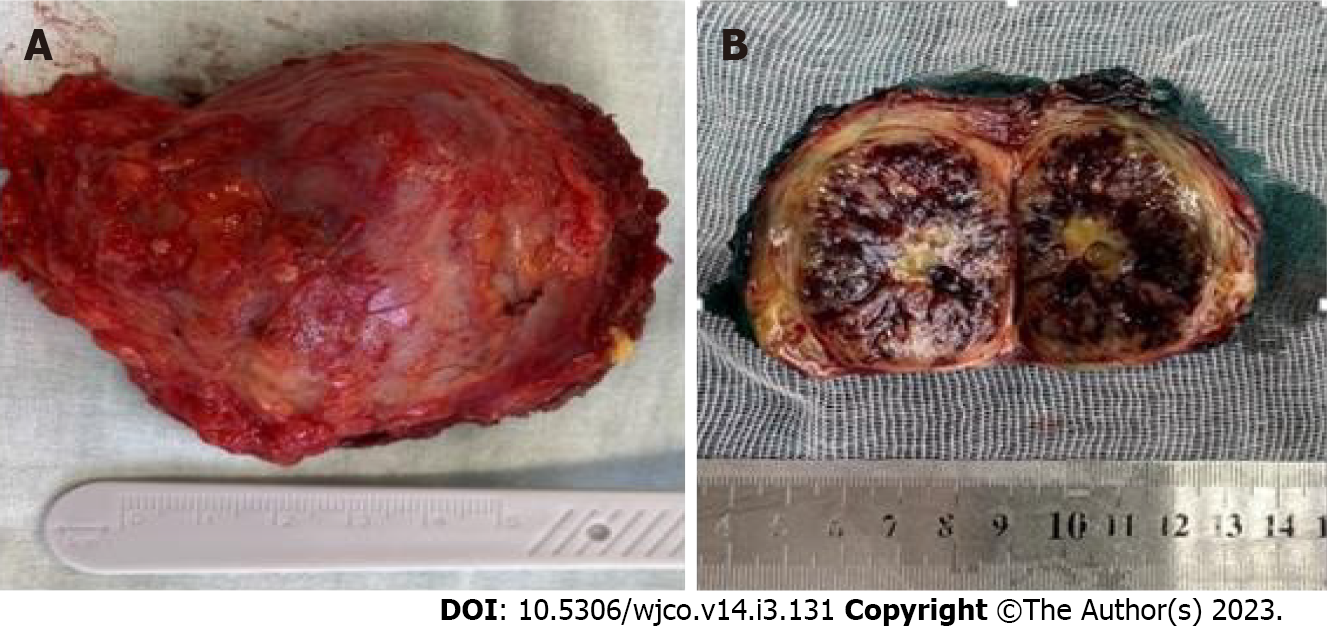
Figure 3 Macroscopic examination.
A, B: Macroscopical size 50 mm, thick fibrous capsule.
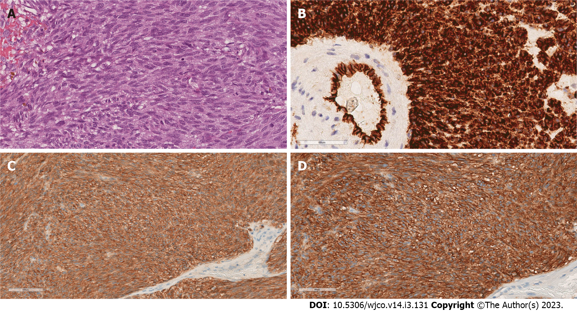
Figure 4 Microscopic examination.
A: Histology, original magnification ×200, hematoxylin-eosin stain; B: Immunohistochemistry original magnification ×200, CD34 positive stain; C: Immunohistochemistry ×200, CD117 positive stain, D: Immunohistochemistry original magnification×200, DOG-1 positive stain.
Figure 5 Treatment timeline.
- Citation: Filonenko D, Karnaukhov N, Kvetenadze G, Zhukova L. Unusual breast metastasis of gastrointestinal stromal tumor: A case report and literature review. World J Clin Oncol 2023; 14(3): 131-137
- URL: https://www.wjgnet.com/2218-4333/full/v14/i3/131.htm
- DOI: https://dx.doi.org/10.5306/wjco.v14.i3.131


