Copyright
©The Author(s) 2022.
World J Clin Oncol. Jun 24, 2022; 13(6): 496-504
Published online Jun 24, 2022. doi: 10.5306/wjco.v13.i6.496
Published online Jun 24, 2022. doi: 10.5306/wjco.v13.i6.496
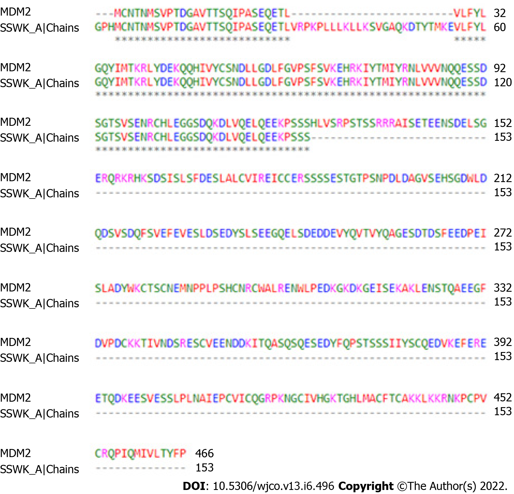
Figure 1 Global alignment between 5SWK chain A (153 residues) and whole mouse double minute 2 (466 residues).
Consensus region is located at position 56 - 153 of 5SWK chain A. The color of the residues represents the chemical characteristic of their side chains. MDM2: Mouse double minute 2.
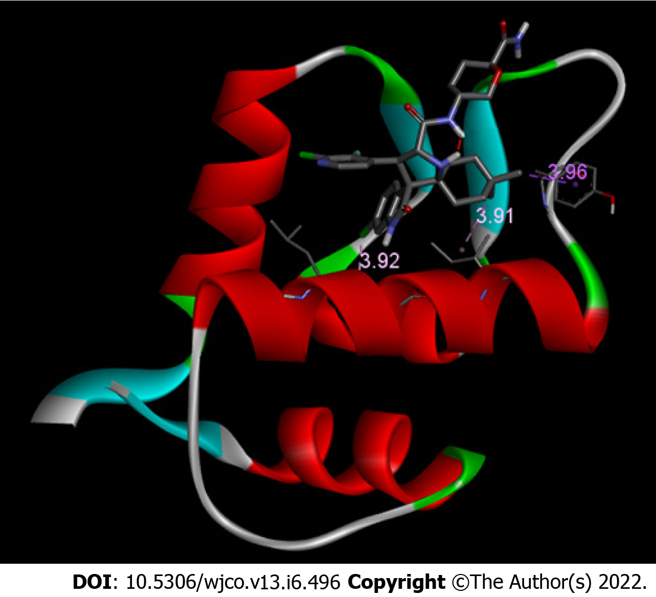
Figure 2 Mouse double minute 2/protonated DS-3032B conformer.
Mouse double minute 2 (MDM2) receptor is shown in ribbon and DS-3032B is shown in sticks. Only LEU 57, ILE 61, and TYR 67 bonds are shown. Bond size in Å.
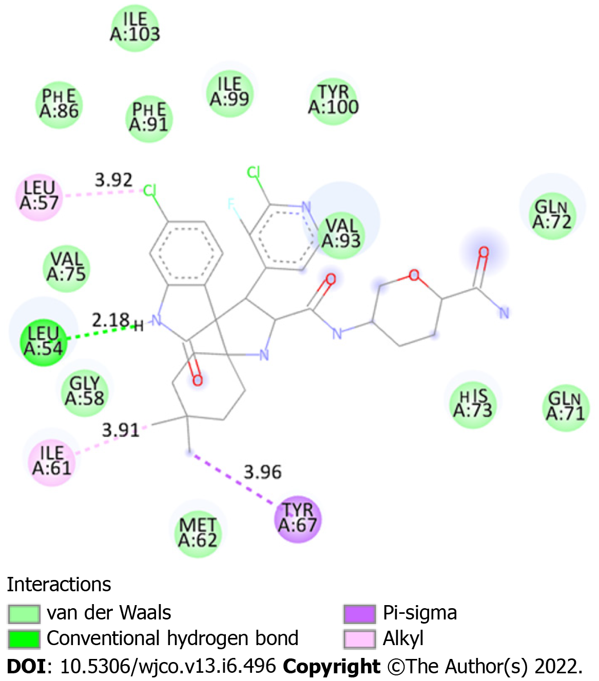
Figure 3 Two-dimensional diagram demonstrating interactions between mouse double minute 2 chain A (5SWK) and protonated DS-3032B.
Light green: Residues involved in Van der Waals interactions. Green: Residue involved in hydrogen bond. Lilac: Residue involved Pi-Sigma interaction. Pink: Residues involved in alkyl interactions. Numbers in spheres indicate the residue position. Bond size are shown (Å).
- Citation: da Mota VHS, Freire de Melo F, de Brito BB, Silva FAFD, Teixeira KN. Molecular docking of DS-3032B, a mouse double minute 2 enzyme antagonist with potential for oncology treatment development. World J Clin Oncol 2022; 13(6): 496-504
- URL: https://www.wjgnet.com/2218-4333/full/v13/i6/496.htm
- DOI: https://dx.doi.org/10.5306/wjco.v13.i6.496


