Copyright
©The Author(s) 2022.
World J Clin Oncol. Apr 24, 2022; 13(4): 303-313
Published online Apr 24, 2022. doi: 10.5306/wjco.v13.i4.303
Published online Apr 24, 2022. doi: 10.5306/wjco.v13.i4.303
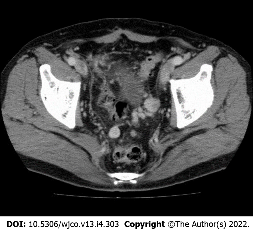
Figure 1 Image of abdominal computed tomography.
Suspected carcinomatosis or sarcomatosis was noted in the pelvis with no evident ascites.
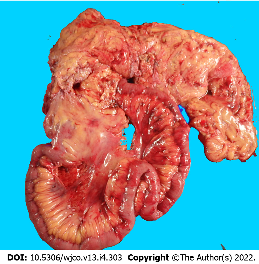
Figure 2 Intestine specimen after extended radical right hemicolectomy.
Multiple whitish tumor nodules seeding over the visceral peritoneum of the partial ileum and colon.
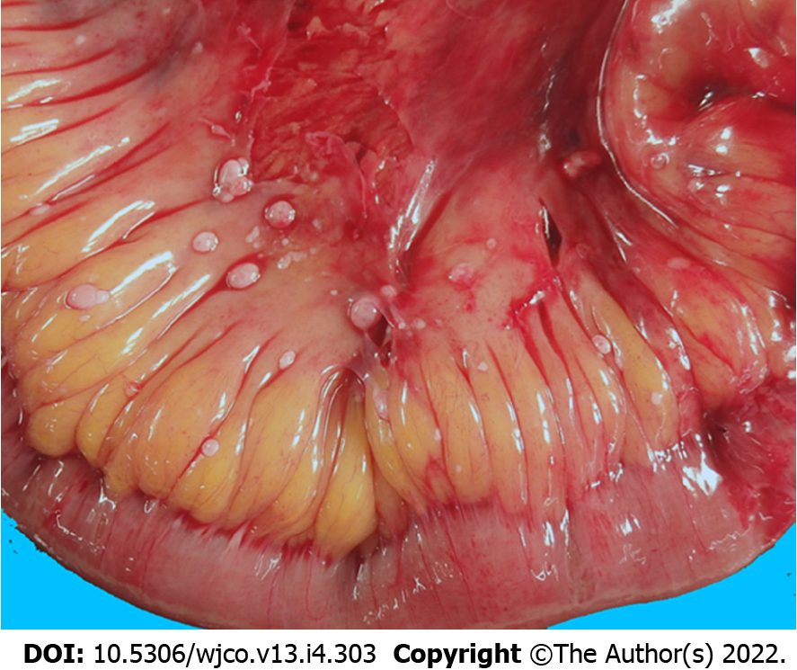
Figure 3 Image of an ileum specimen.
Multiple whitish tumor nodules seeding over the visceral peritoneum of the distal ileum.
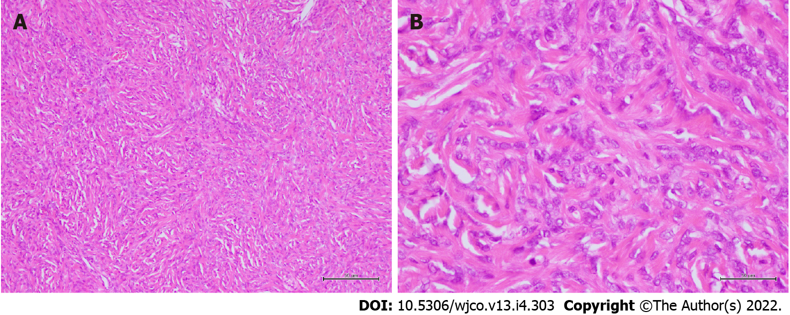
Figure 4 Microscopic features.
The heterogeneous cell population comprised of mostly spindle cells with fibrous collagen proliferation as well as various other cell populations exhibiting patternless or storiform growth. No tumor necrosis, nuclear polymorphism, or cellular atypia was noted. The nuclear mitotic index was 4 mitoses/50 high-power fields (A: × 100 original magnification; B: × 400 original magnification).
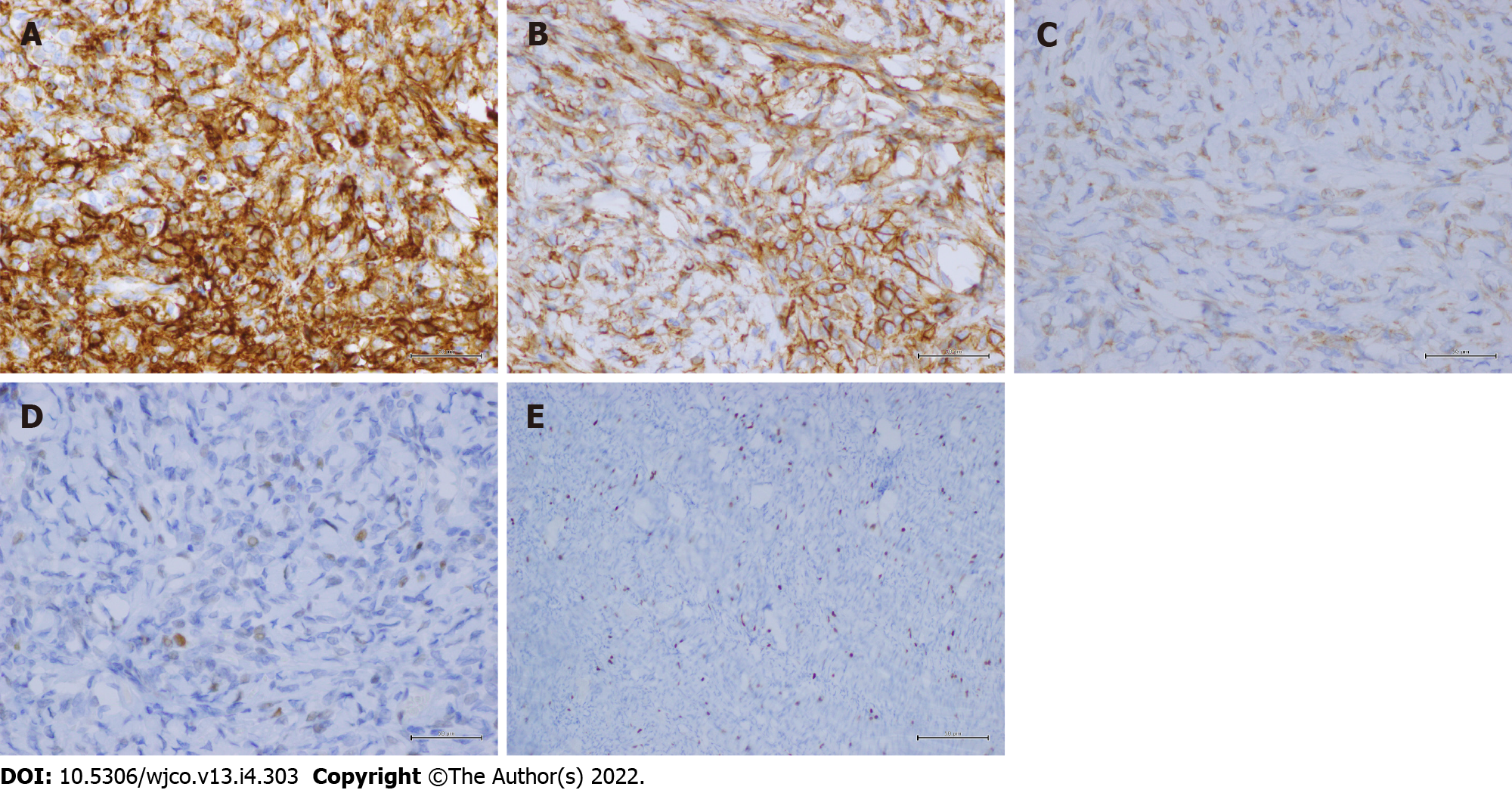
Figure 5 Immunohistochemical staining.
A-E: The lesion showed diffuse strong staining for CD34 (× 400 original magnification) (A); CD99 (× 400 original magnification) (B); Bcl-2 (× 400 original magnification) (C); mildly positive immunoreactivity for p53 (× 400 original magnification) (D); and a Ki-67 mitotic proliferative index of approximately 5% (× 100 original magnification) (E).
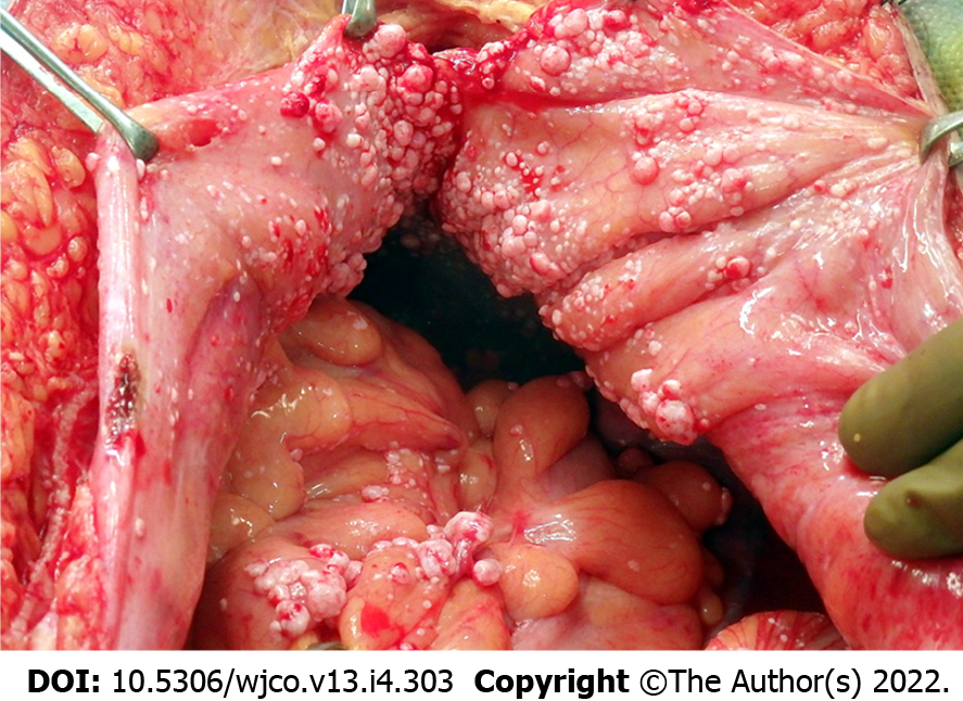
Figure 6 Image of the pelvis during operation.
Multiple whitish nodules were noted to be seeded over the parietal peritoneum and visceral peritoneum of the partial ileum and colon as well as the urinary bladder, with no ascites in the pelvis.
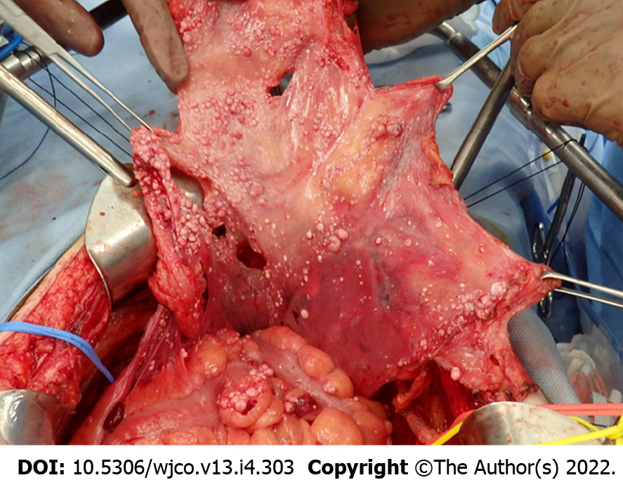
Figure 7 Peritonectomy during cytoreductive surgery.
Total anterior parietal peritonectomy, bilateral subphrenic peritonectomy, and complete pelvic peritonectomy (including the visceral peritoneum covering the urinary bladder) were performed.
- Citation: Chiu CC, Ishibashi H, Wakama S, Liu Y, Hao Y, Hung CM, Lee PH, Rau KM, Lee HM, Yonemura Y. Mesentery solitary fibrous tumor with postoperative recurrence and sarcomatosis: A case report and review of literature. World J Clin Oncol 2022; 13(4): 303-313
- URL: https://www.wjgnet.com/2218-4333/full/v13/i4/303.htm
- DOI: https://dx.doi.org/10.5306/wjco.v13.i4.303


