Copyright
©The Author(s) 2022.
World J Clin Oncol. Oct 24, 2022; 13(10): 853-860
Published online Oct 24, 2022. doi: 10.5306/wjco.v13.i10.853
Published online Oct 24, 2022. doi: 10.5306/wjco.v13.i10.853
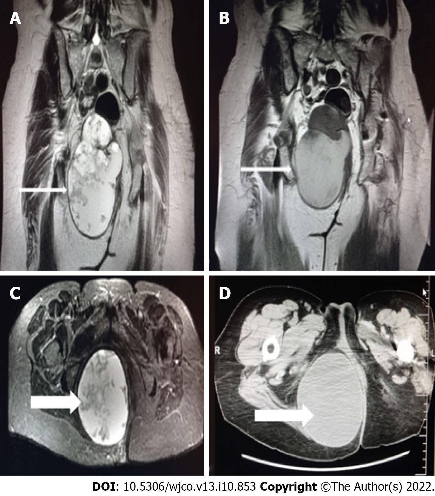
Figure 1 Magnetic resonance imaging.
A and C: Coronal and axial planes of the mass with smooth borders, lobed on the upper side with a beak sign. Cystic and solid elements, septa, and haemorrhagic and protein elements. It absorbs paramagnetic substance; B and D: Computed tomography scan - Coronal and axial planes of the mass. Differential diagnosis of tail gut cyst or cystic teratoma (arrows).
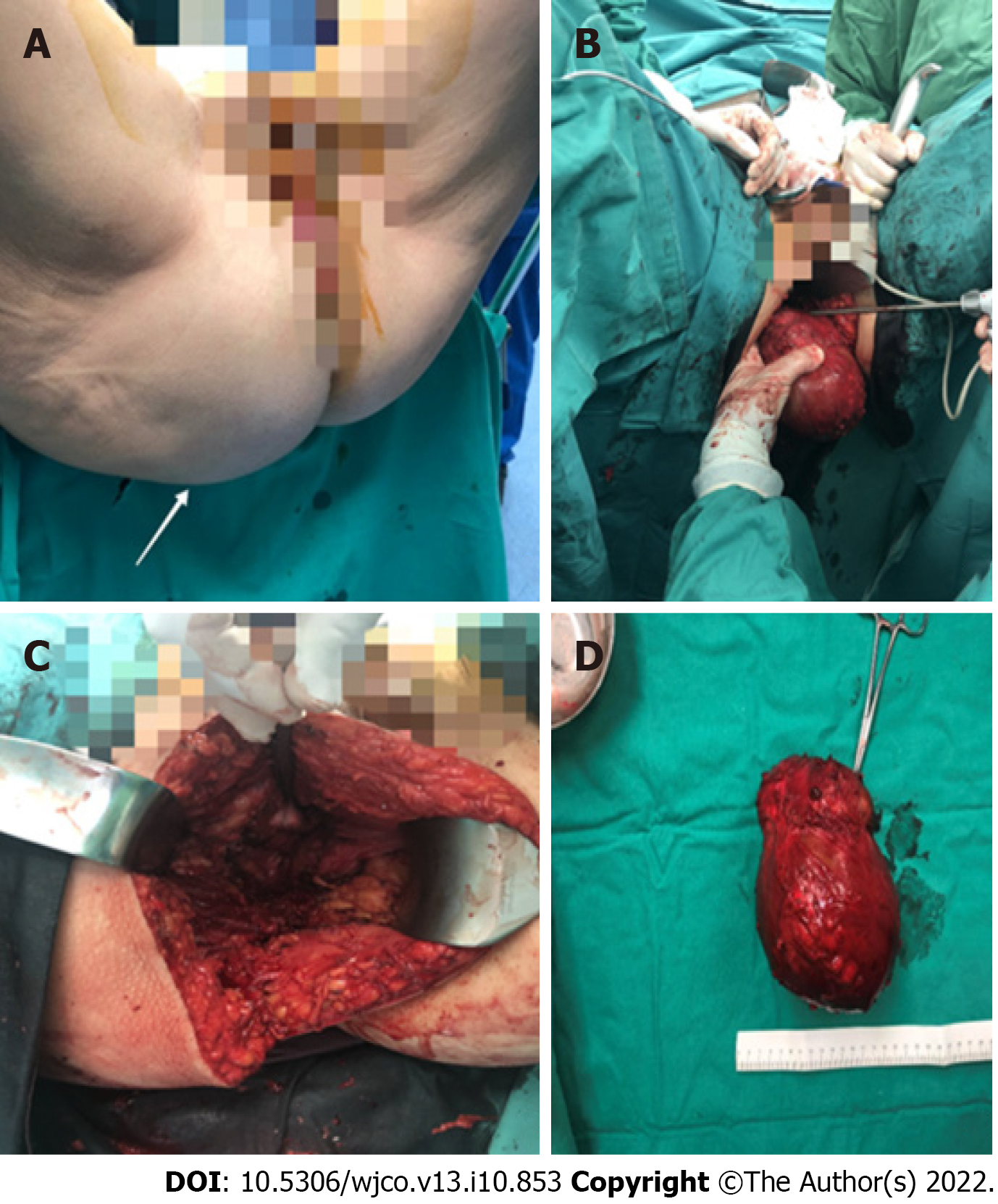
Figure 2 Patient underwent extensive surgical resection of the lesion through the right buttock.
A: Preoperative view of the mass (arrow); B and C: Extensive surgical resection of the lesion through the right buttock; D: Removed mass.
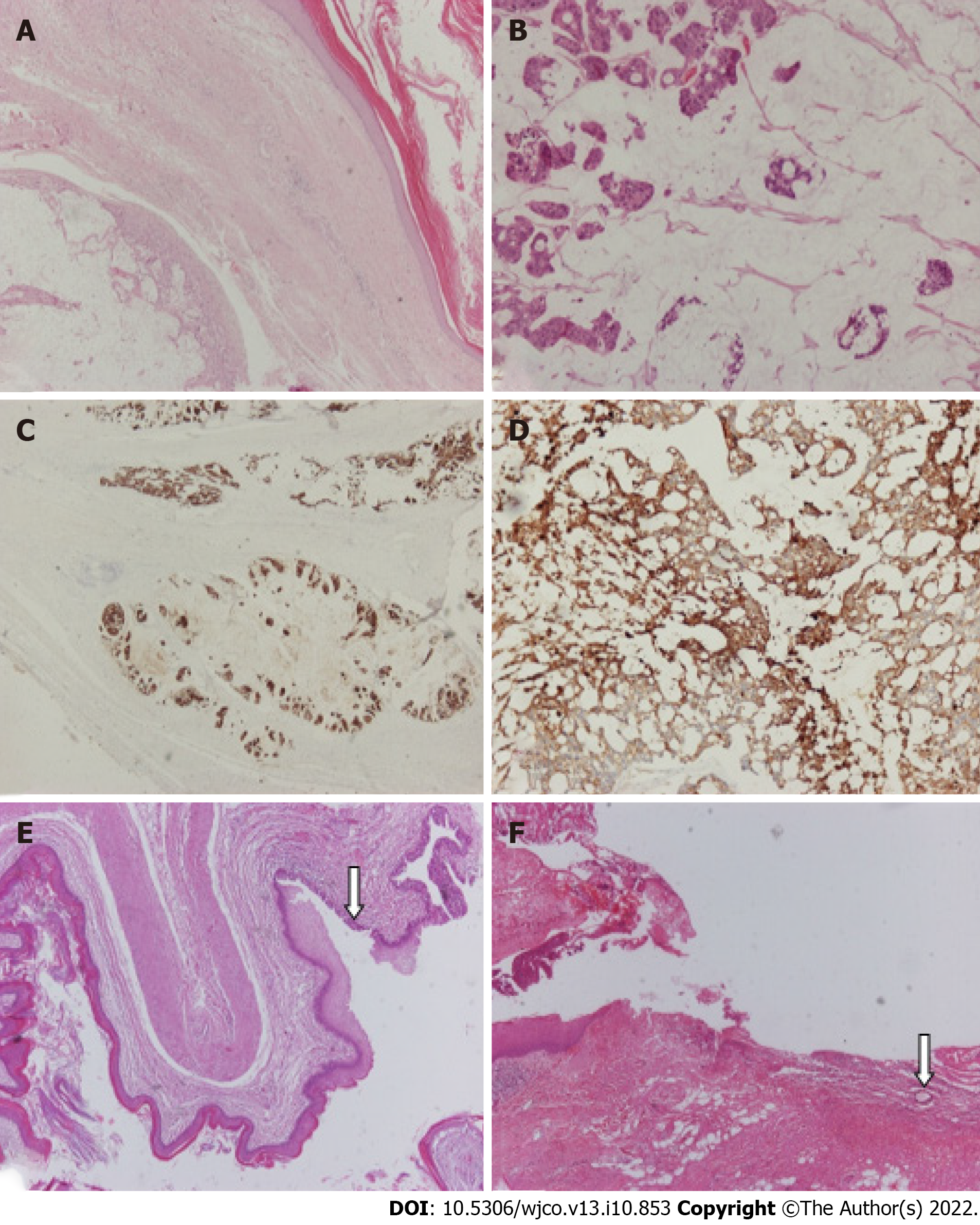
Figure 3 Microscopic examination confirmed the presence of a cystic mass that comprised thick fibrous bands that divided it into three cystic spaces, the largest of which corresponded to mucinous adenocarcinoma.
A: Fibrous tissue separates two cystic spaces, one benign lined by keratinized squamous cell epithelium and the other corresponding to mucinous adenocarcinoma; B-D: Higher magnification of mucinous adenocarcinoma that is immunohistochemically positive (IHC-positive) for antibodies CK20 and CK7; E and F: A smaller cystic space with fibromuscular wall lined by keratinized squamous epithelium and partially by pseudostratified ciliated columnar epithelium (arrow) is observed. Section of the pilonidal cyst (arrow: Hair shaft). [A: Hematoxylin-eosin staining (HE), magnification × 40; B: HE × 100; C: IHC × 20; D: IHC × 100; E and F: HE × 40].
- Citation: Malliou P, Syrnioti A, Koletsa T, Karlafti E, Karakatsanis A, Raptou G, Apostolidis S, Michalopoulos A, Paramythiotis D. Mucinous adenocarcinoma arising from a tailgut cyst: A case report. World J Clin Oncol 2022; 13(10): 853-860
- URL: https://www.wjgnet.com/2218-4333/full/v13/i10/853.htm
- DOI: https://dx.doi.org/10.5306/wjco.v13.i10.853


