Copyright
©The Author(s) 2021.
World J Clin Oncol. Sep 24, 2021; 12(9): 808-822
Published online Sep 24, 2021. doi: 10.5306/wjco.v12.i9.808
Published online Sep 24, 2021. doi: 10.5306/wjco.v12.i9.808
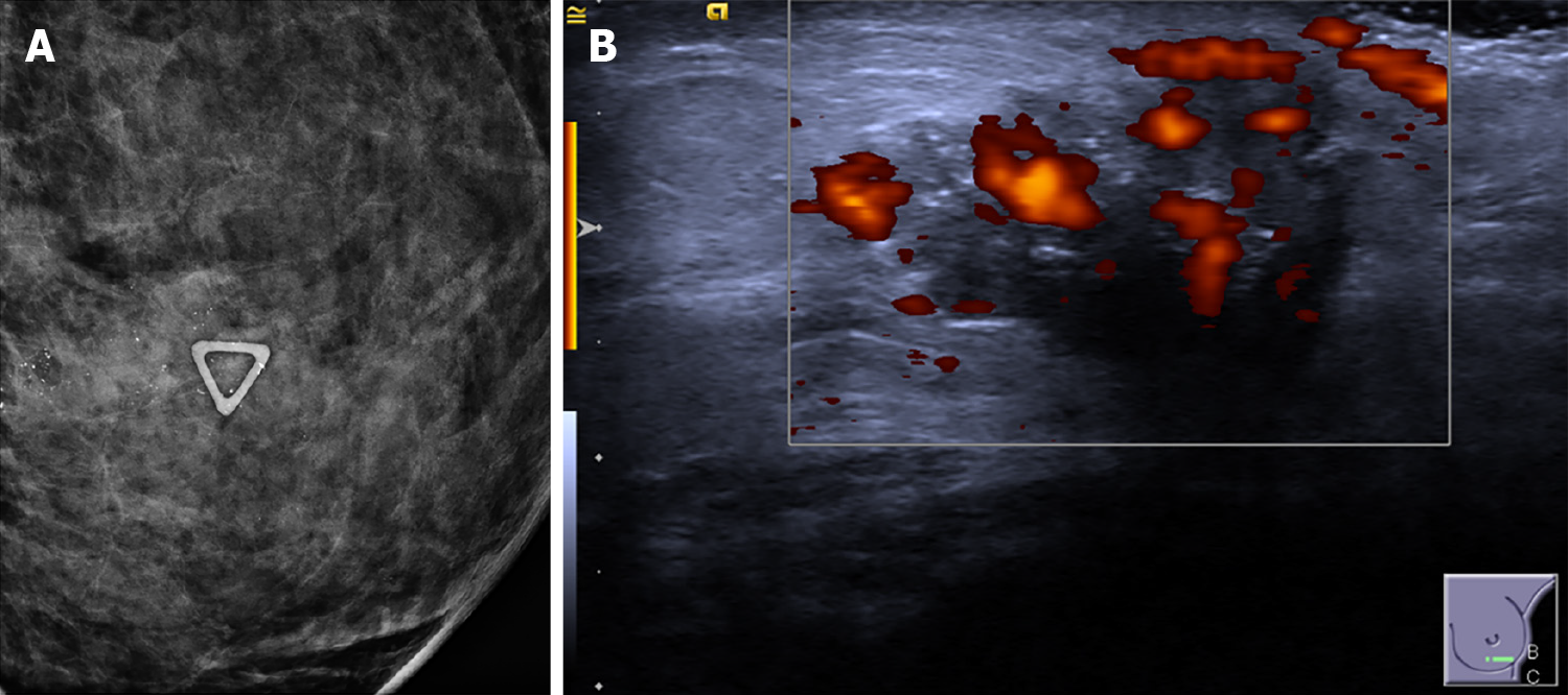
Figure 1 HER2-enriched invasive cancer.
A: High-risk microcalcifications on mammogram of the left breast are seen within the palpable mass (denoted by triangular skin marker); B: Irregular hypoechoic mass showing increased internal vascularity (Adler Index Grade III) and internal echogenic foci representing microcalcifications.
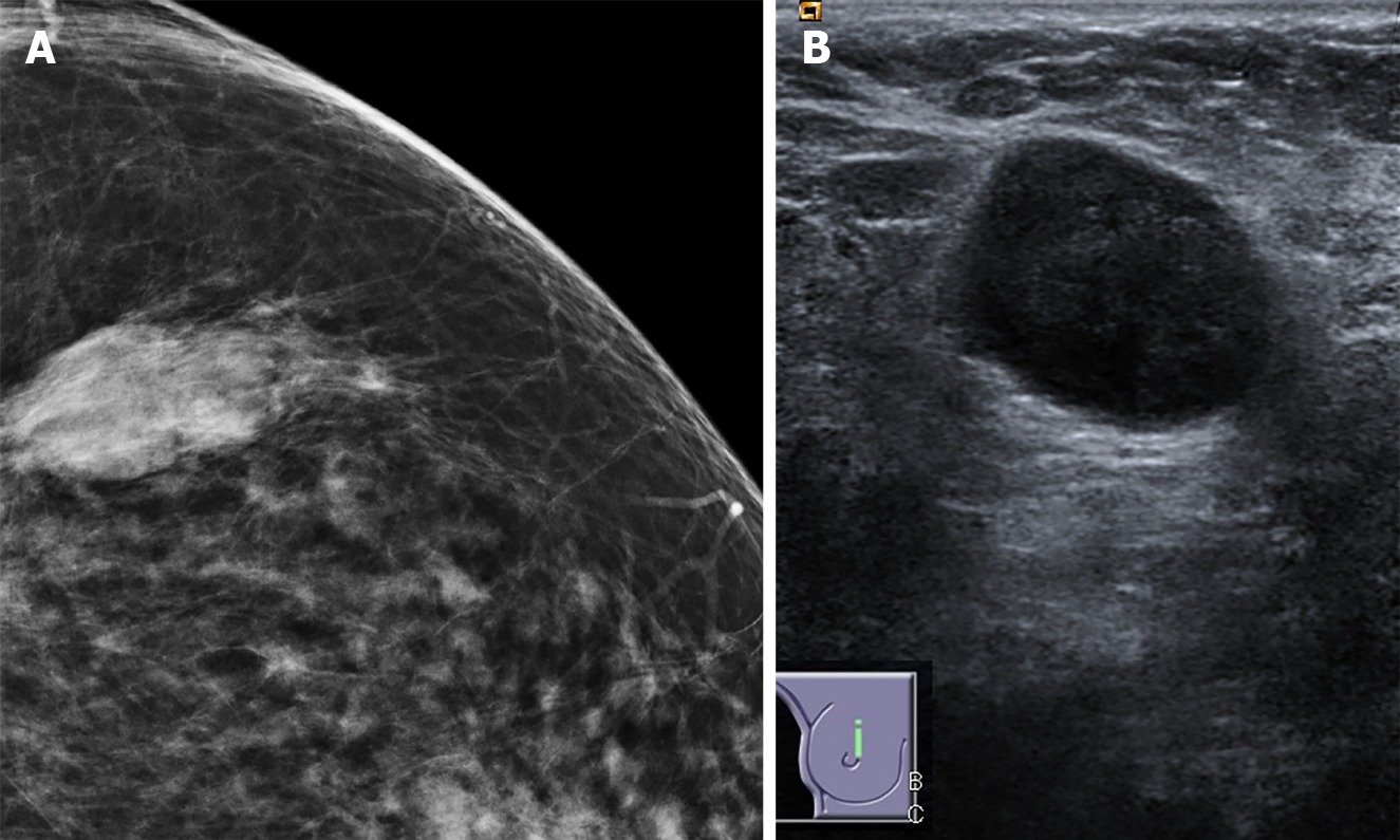
Figure 2 Triple negative breast cancer.
A: Circumscribed mass on mammogram; B: Circumscribed hypoechoic mass with posterior acoustic enhancement on ultrasound.
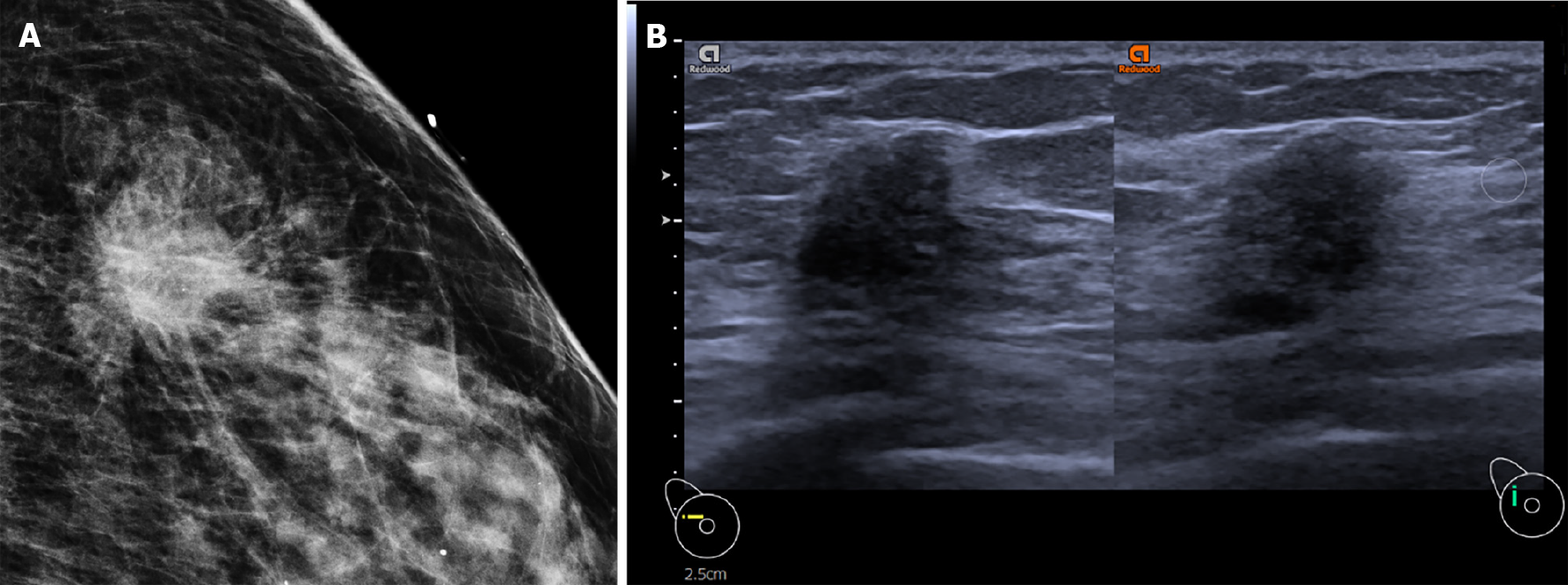
Figure 3 Luminal type invasive cancer.
A: Spiculated mass on mammogram; B: Irregular hypoechoic mass with spiculated margin and posterior acoustic shadowing.
- Citation: Ian TWM, Tan EY, Chotai N. Role of mammogram and ultrasound imaging in predicting breast cancer subtypes in screening and symptomatic patients. World J Clin Oncol 2021; 12(9): 808-822
- URL: https://www.wjgnet.com/2218-4333/full/v12/i9/808.htm
- DOI: https://dx.doi.org/10.5306/wjco.v12.i9.808


