Copyright
©The Author(s) 2021.
World J Clin Oncol. Apr 24, 2021; 12(4): 238-248
Published online Apr 24, 2021. doi: 10.5306/wjco.v12.i4.238
Published online Apr 24, 2021. doi: 10.5306/wjco.v12.i4.238
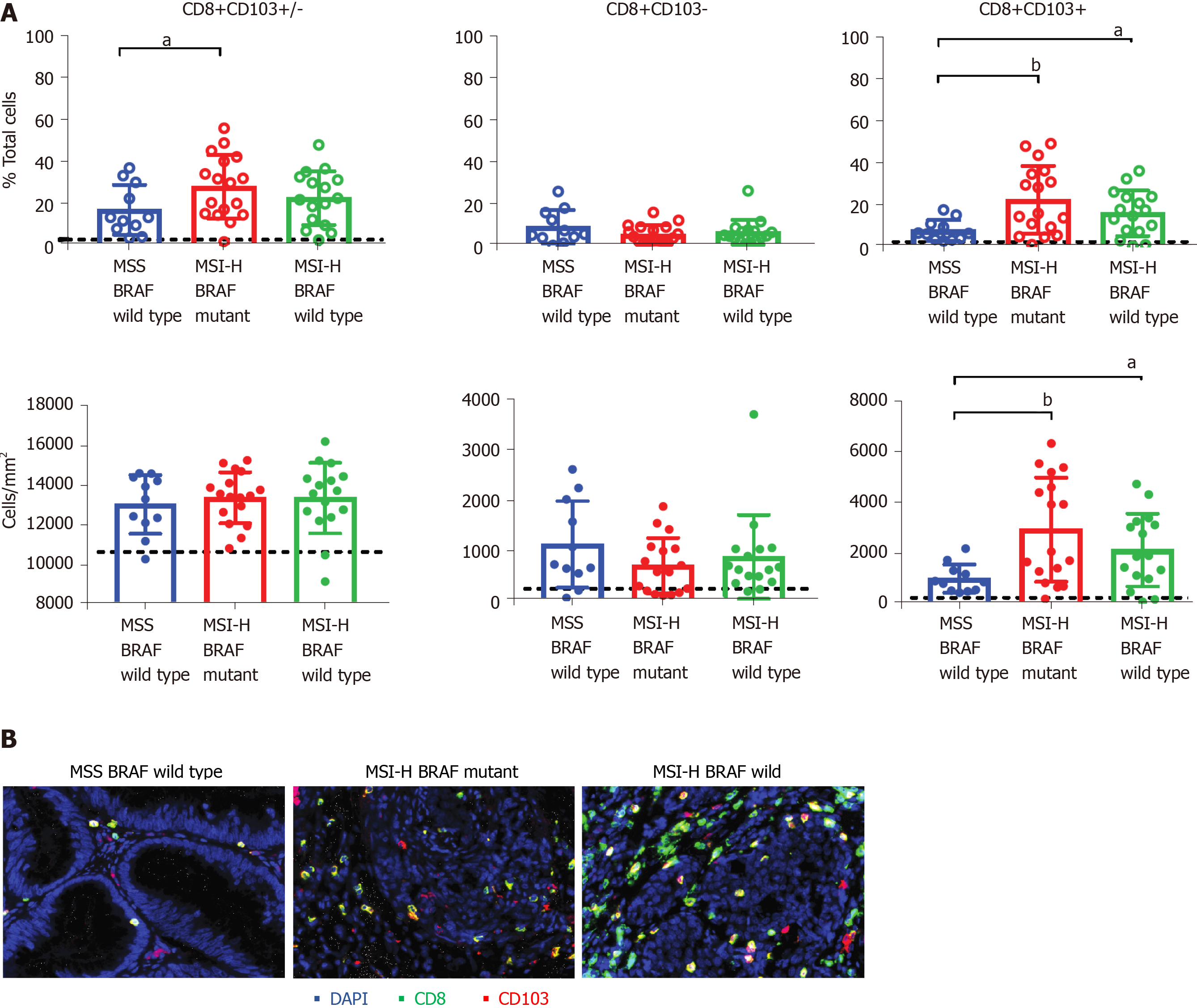
Figure 1 Tissue resident memory T cells are significantly increased in microsatellite instability high tumours.
A: Quantitative immunohistochemistry staining of tumour specimen show the proportion (top panels) and densities (bottom panels) of total CD8+ T cells (left panels), CD103-CD8+ (non-TRM) T cells (middle panels) and CD103+CD8+ resident memory (TRM) T cells (right panels) in CRC tumours with microsatellite stable BRAF wildtype and microsatellite instability (MSI) high BRAF mutants and MSI high BRAF wildtype groups. Figure shows the mean and SD of n = 11-17 samples and the statistical differences were calculated using unpaired and non-parametric Mann-Whitney test T test (aP < 0.05; bP < 0.001). Black line shows the levels determined from normal healthy colorectal tissues (n = 1); B: Representative tissue staining shows the staining for CD8 (green) and CD103 (red) and DAPI (blue) in the three groups of patients. MSS: Microsatellite stable; MSI-H: High microsatellite instability.
Figure 2 Programmed cell death protein-1 expression on CD8+ T cells is not impacted by microsatellite instability or BRAF status.
Quantitative immunohistochemistry staining was used to determine the expression of programmed cell death protein-1 (PD-1) on CD8+ T cell populations. Figure shows the proportion of total CD8+ T cells (CD8+CD103+/-) (left panel), CD103-CD8+ (non-TRM) T cells (middle panel) and CD103+CD8+ TRM cells (right panel) expressing PD-1. Figure shows the mean and SD for 5-6 samples/group. PD-1: Programmed death 1; MSS: Microsatellite stable; MSI-H: High microsatellite instability.
- Citation: Toh JWT, Ferguson AL, Spring KJ, Mahajan H, Palendira U. Cytotoxic CD8+ T cells and tissue resident memory cells in colorectal cancer based on microsatellite instability and BRAF status. World J Clin Oncol 2021; 12(4): 238-248
- URL: https://www.wjgnet.com/2218-4333/full/v12/i4/238.htm
- DOI: https://dx.doi.org/10.5306/wjco.v12.i4.238


