Copyright
©The Author(s) 2020.
World J Clin Oncol. Mar 24, 2020; 11(3): 121-135
Published online Mar 24, 2020. doi: 10.5306/wjco.v11.i3.121
Published online Mar 24, 2020. doi: 10.5306/wjco.v11.i3.121
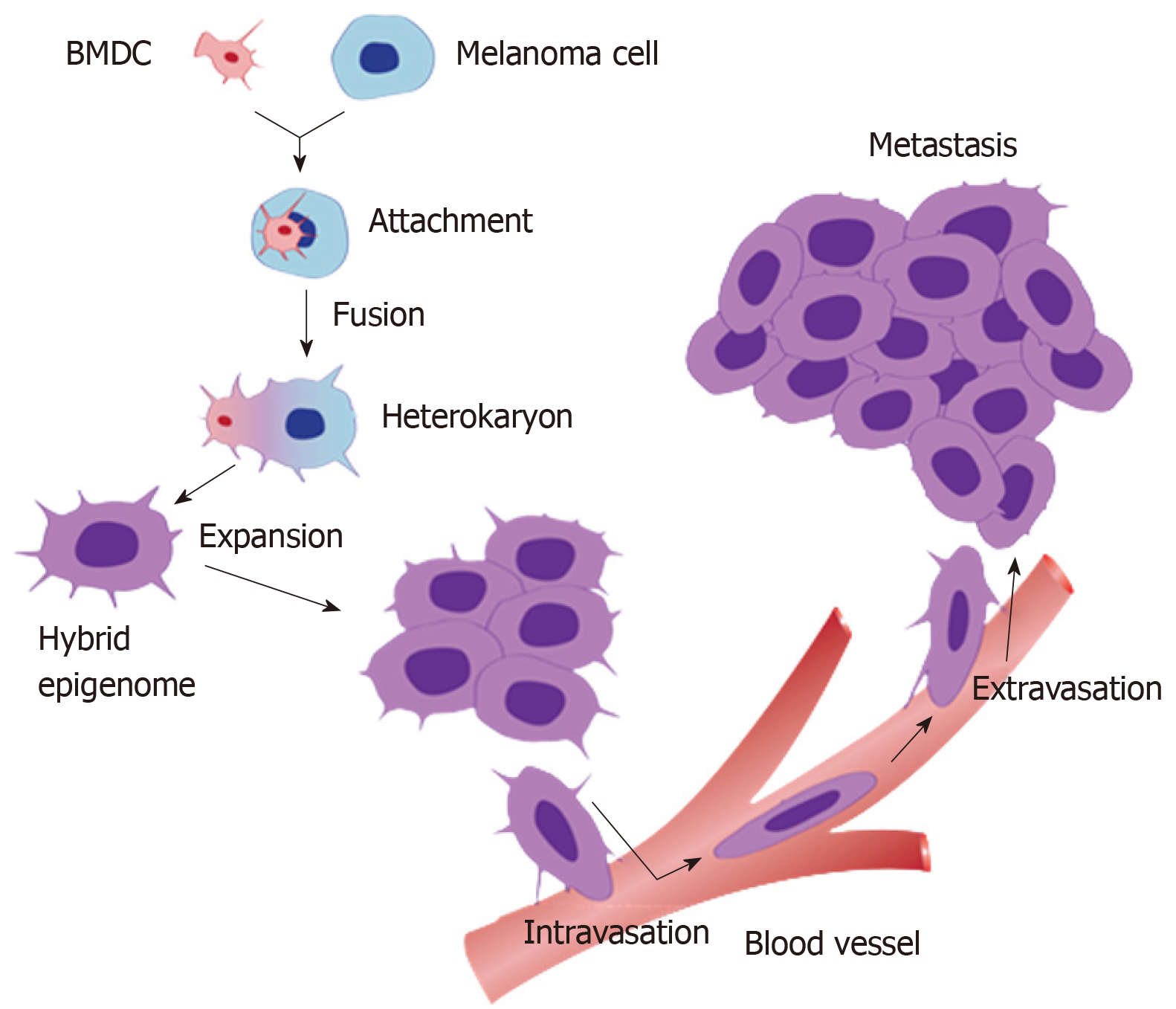
Figure 1 A schematic diagram of the process of cell fusion, hybrid formation and metastasis.
A motile bone marrow derived cells (red) such as a macrophage or stem cell is drawn to a cancer cell (blue). The outer cell membranes of the two cells become attached. Fusion occurs with the formation of a bi-nucleated heterokaryon having a nucleus from each of the fusion partners. The heterokaryon goes through genomic hybridization creating a cancer cell-bone marrow derived cells hybrid with co-expressed epigenomes, conferring deregulated cell division and metastatic competence to the hybrid. BMDC: Bone marrow derived cell.
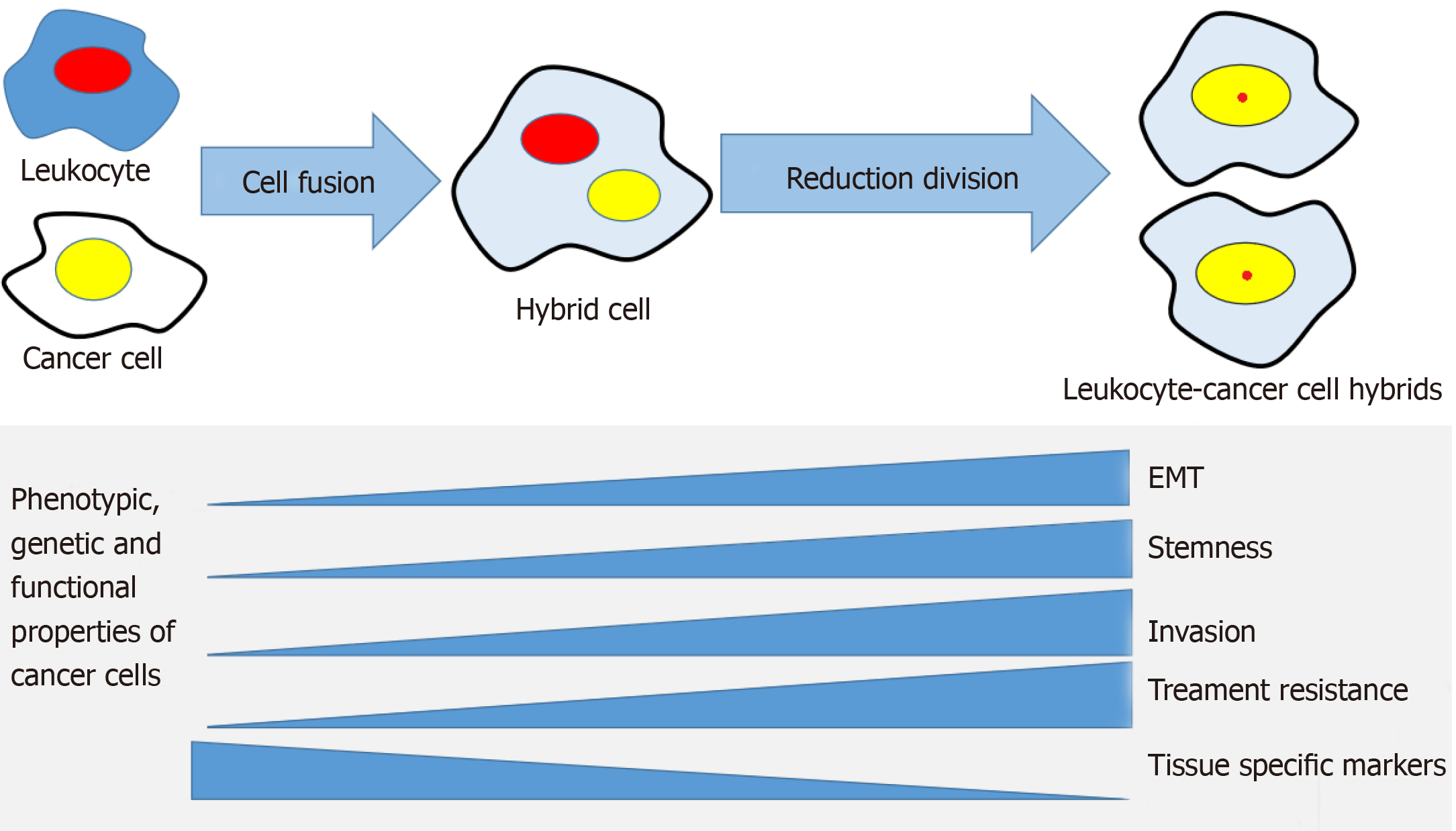
Figure 2 The cell fusion theory in relation to cancer progression mechanisms.
A cancer cell and a leukocyte form a hybrid that acquires genetic, phenotypic and functional properties from both maternal cells. The hybrid cells develop properties associated with cancer metastasis, such as epithelial mesenchymal transition, stemness, invasiveness, treatment resistance and may lose some of the maternal cancer cell's tissue-specific phenotype. EMT: Epithelial to mesenchymal transition.
- Citation: Shabo I, Svanvik J, Lindström A, Lechertier T, Trabulo S, Hulit J, Sparey T, Pawelek J. Roles of cell fusion, hybridization and polyploid cell formation in cancer metastasis. World J Clin Oncol 2020; 11(3): 121-135
- URL: https://www.wjgnet.com/2218-4333/full/v11/i3/121.htm
- DOI: https://dx.doi.org/10.5306/wjco.v11.i3.121


