Copyright
©The Author(s) 2020.
World J Clin Oncol. Nov 24, 2020; 11(11): 868-889
Published online Nov 24, 2020. doi: 10.5306/wjco.v11.i11.868
Published online Nov 24, 2020. doi: 10.5306/wjco.v11.i11.868
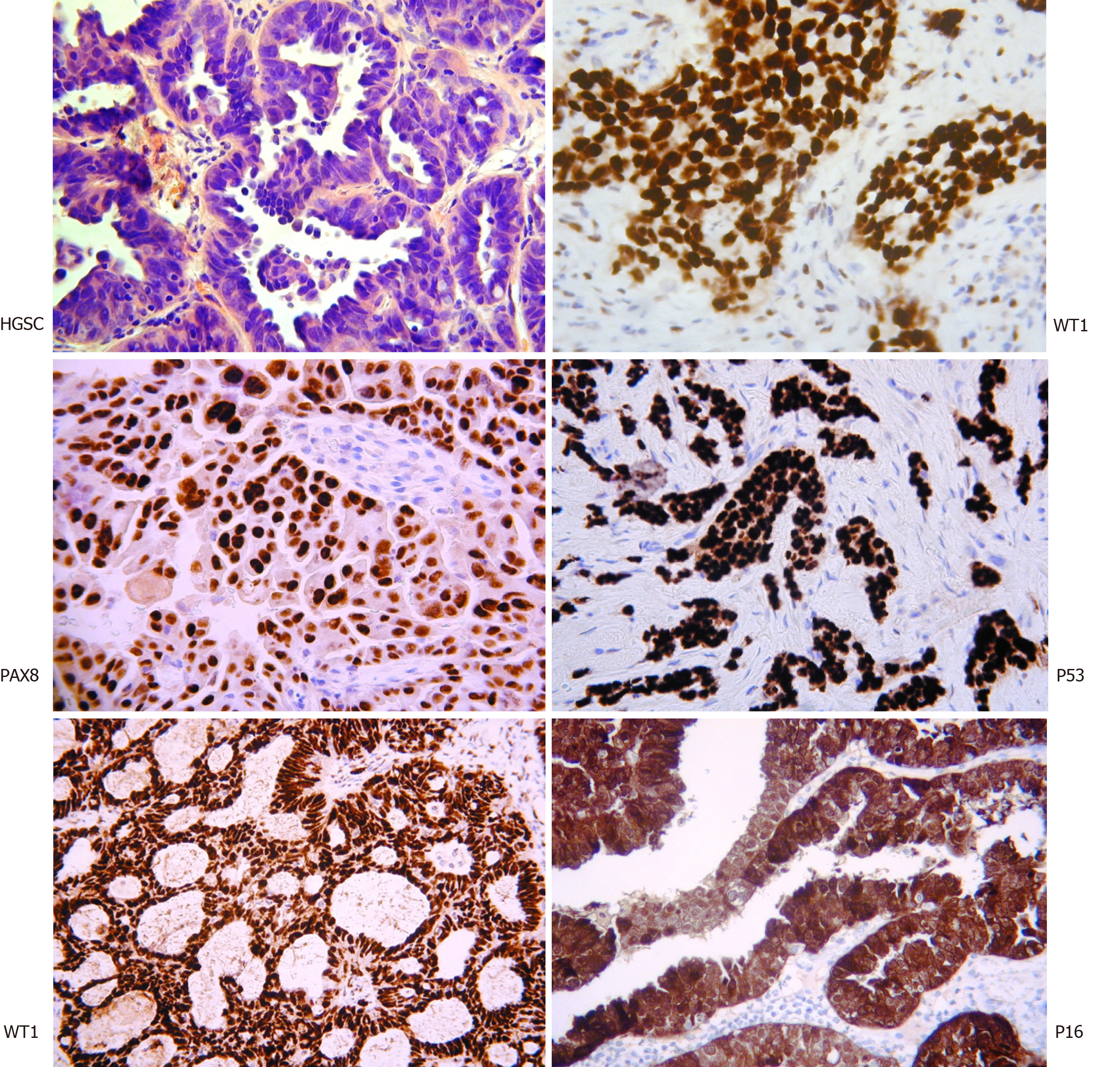
Figure 1 Histopathological assessment of high-grade serous carcinoma.
The classical appearance on hematoxylin and eosin with intermediate sized tumor cells, marked nuclear atypia, and necrotic areas. Immunostaining with WT1, PAX8, P16, and P53 assist with the diagnosis. Interestingly WT1 staining helps discriminate between high-grade serous carcinoma and pseudo-endometrioid (bottom left) (Original figure, images courtesy of Professor McCluggage WG).
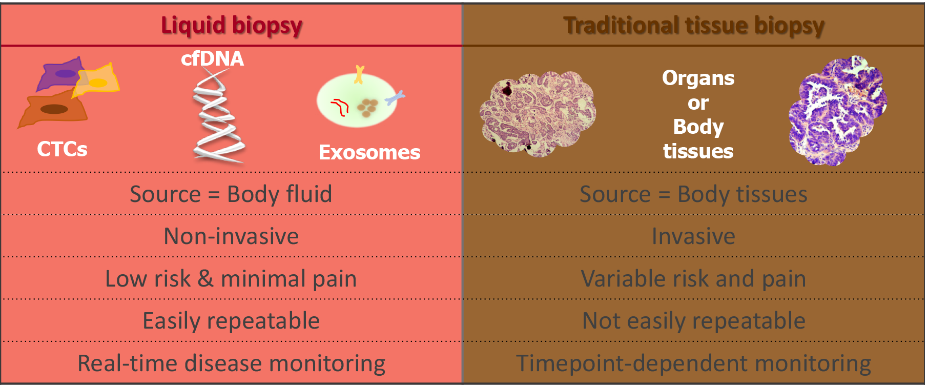
Figure 2 The difference between liquid and traditional tissue biopsy (Original figure).
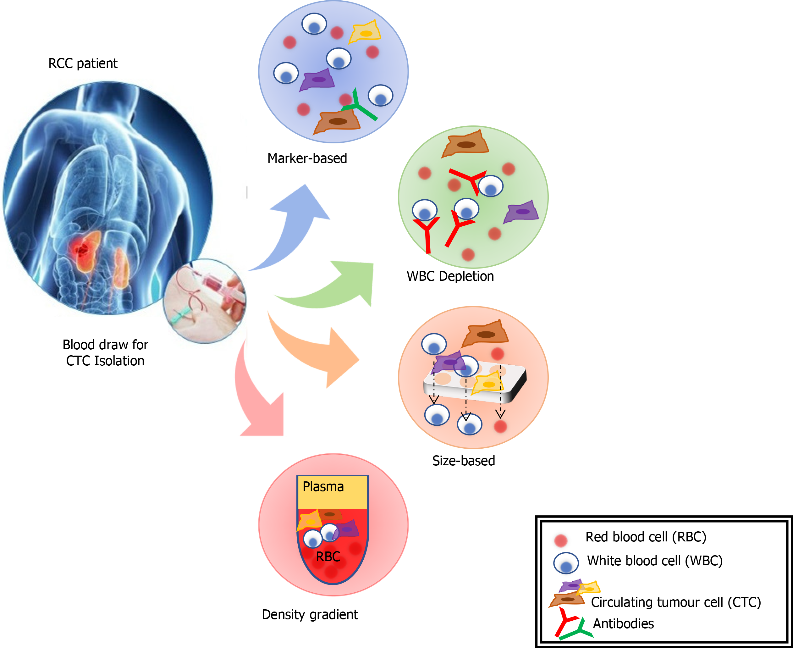
Figure 3 The extraction of circulating tumor cells can be undertaken by a range of methods (reproduced from Broncy et al[166] 2018 under CC BY-NC 4.
0). RCC: Renal cell carcinoma.
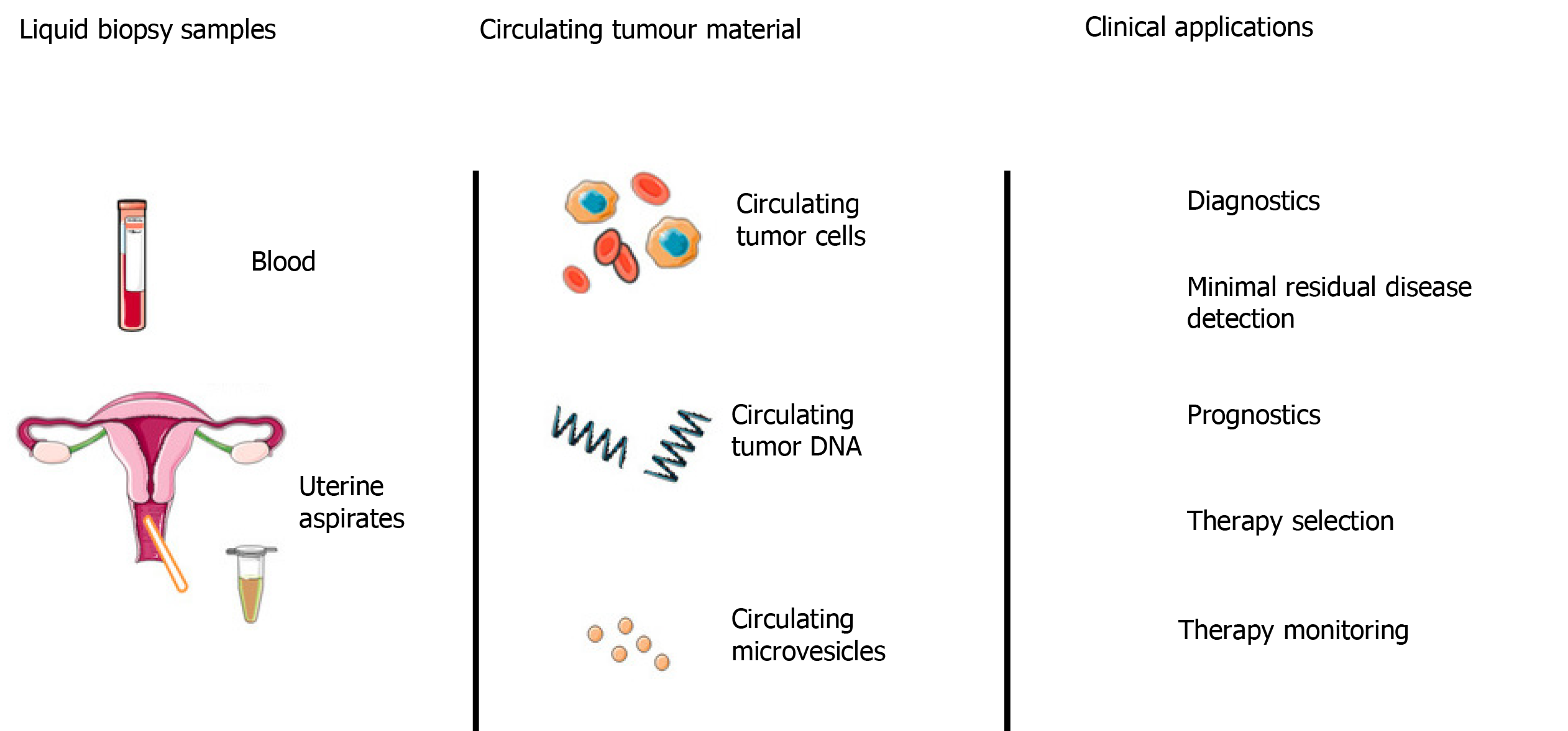
Figure 4 Potential liquid biopsy sources (blood, uterine/cervical aspirates) and downstream clinical applications within epithelial ovarian cancer (reproduced from Muinelo-Romay et al[167] 2018 under CC BY-NC 4.
0).
- Citation: Feeney L, Harley IJ, McCluggage WG, Mullan PB, Beirne JP. Liquid biopsy in ovarian cancer: Catching the silent killer before it strikes. World J Clin Oncol 2020; 11(11): 868-889
- URL: https://www.wjgnet.com/2218-4333/full/v11/i11/868.htm
- DOI: https://dx.doi.org/10.5306/wjco.v11.i11.868


