Copyright
©The Author(s) 2020.
World J Clin Oncol. Oct 24, 2020; 11(10): 844-853
Published online Oct 24, 2020. doi: 10.5306/wjco.v11.i10.844
Published online Oct 24, 2020. doi: 10.5306/wjco.v11.i10.844
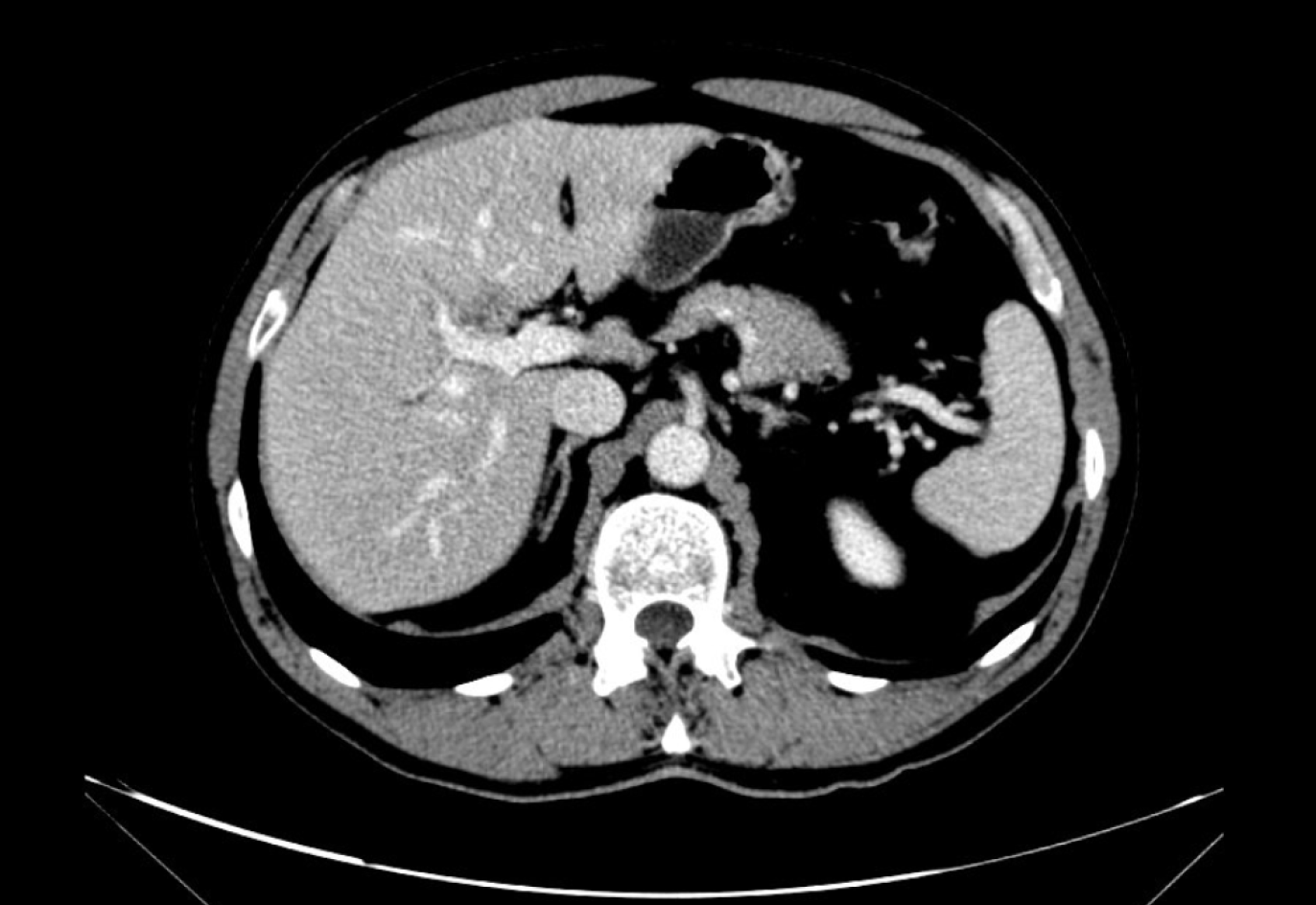
Figure 1 Computed tomography scan performed in December 2016.
A 20 mm hypodense intrahepatic mass at the right lobe, possible cholangiocarcinoma, was reported.
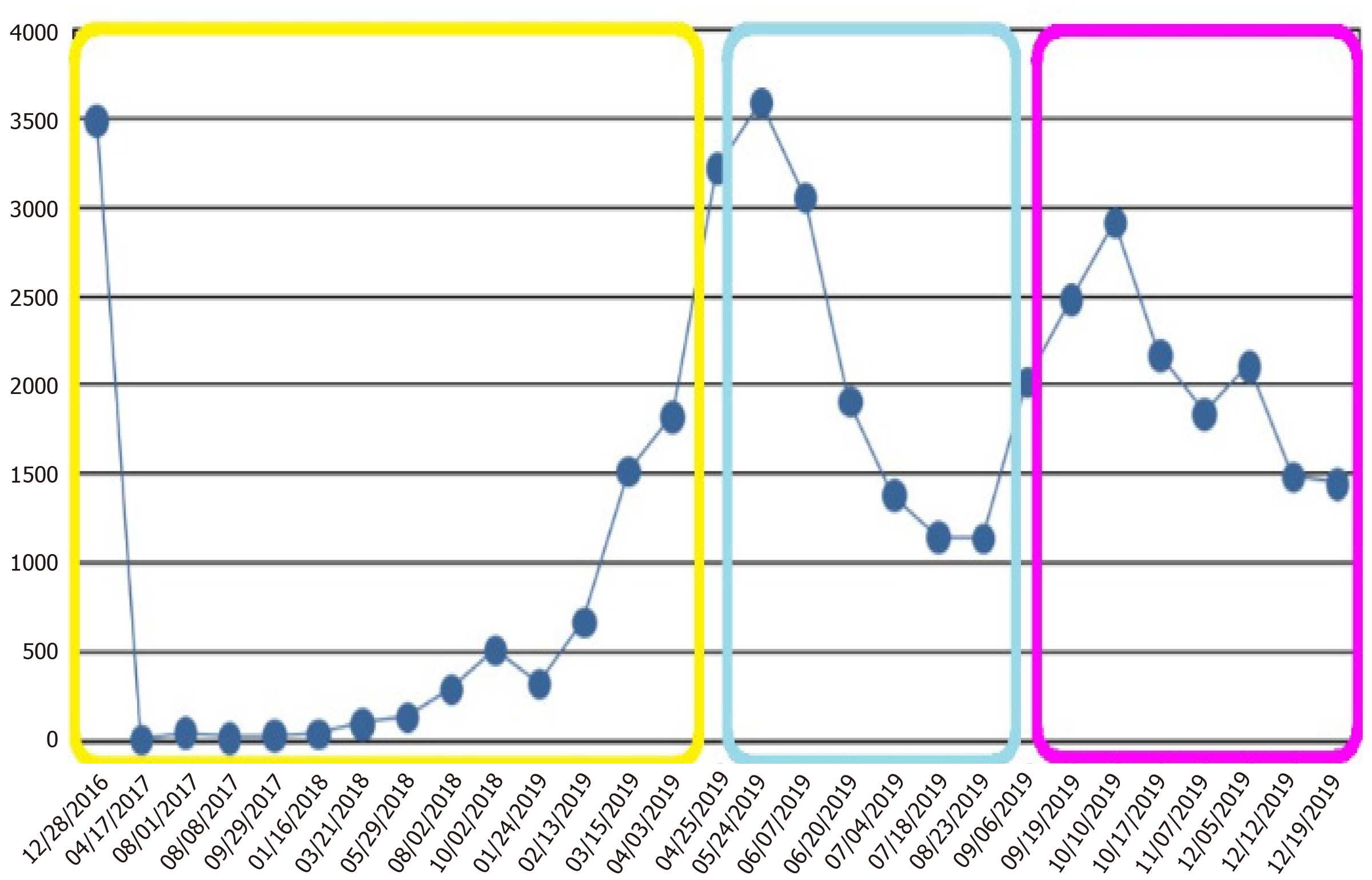
Figure 2 Evolution of CA19-9 blood levels (IU/mL).
Yellow: CA19-9 levels of localized cholangiocarcinoma at diagnosis, after surgery, and during follow-up; Cyan: CA19-9 levels during FOLFOX treatment; Magenta: CA19-9 levels during albumin-bound paclitaxel treatment.
Figure 3 Positron emission tomography-computed tomography performed in April 2019.
Cholangiocarcinoma relapse in segment IVa liver node (before FOLFOX6 treatment).
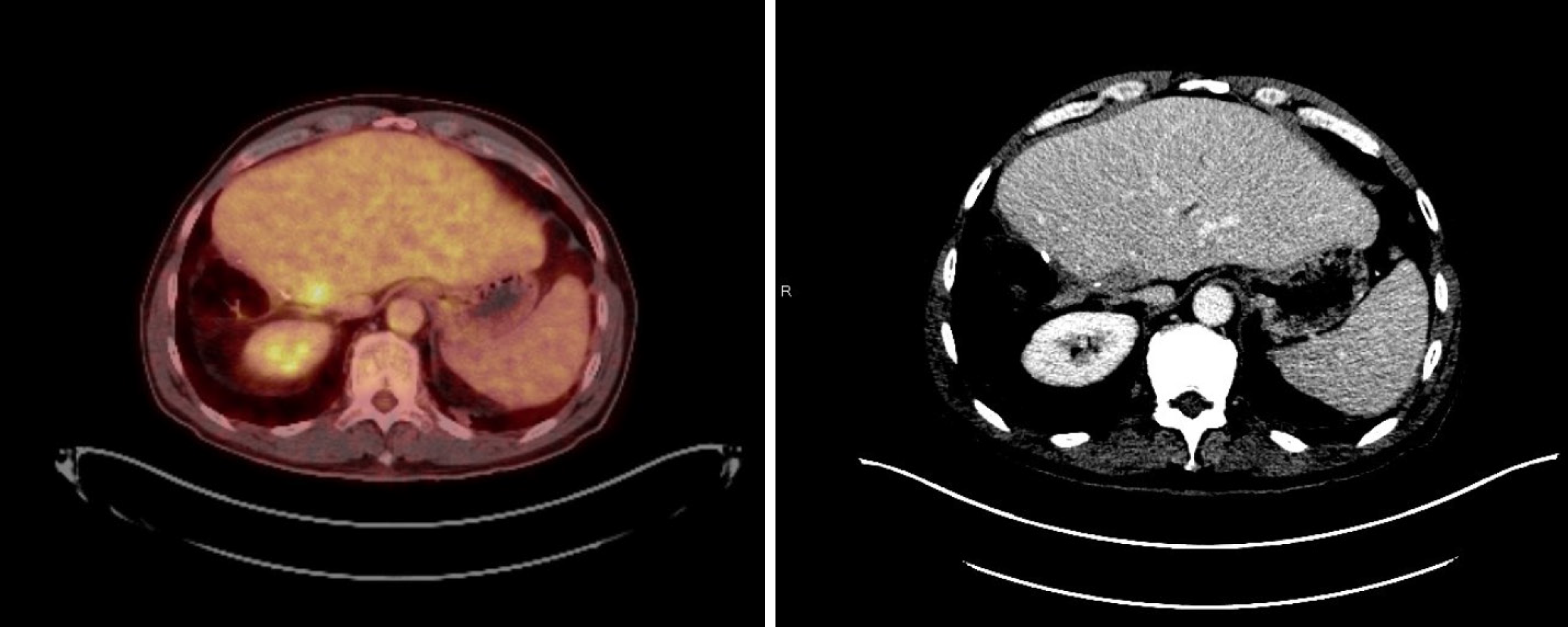
Figure 4 Computed tomography scans performed in July 2019 (left, after seven cycles of FOLFOX6 treatment, where a complete response of disease in segment IV liver is shown) and April 2019 (right, before FOLFOX6 treatment, where segment IV liver node metastasis is seen).
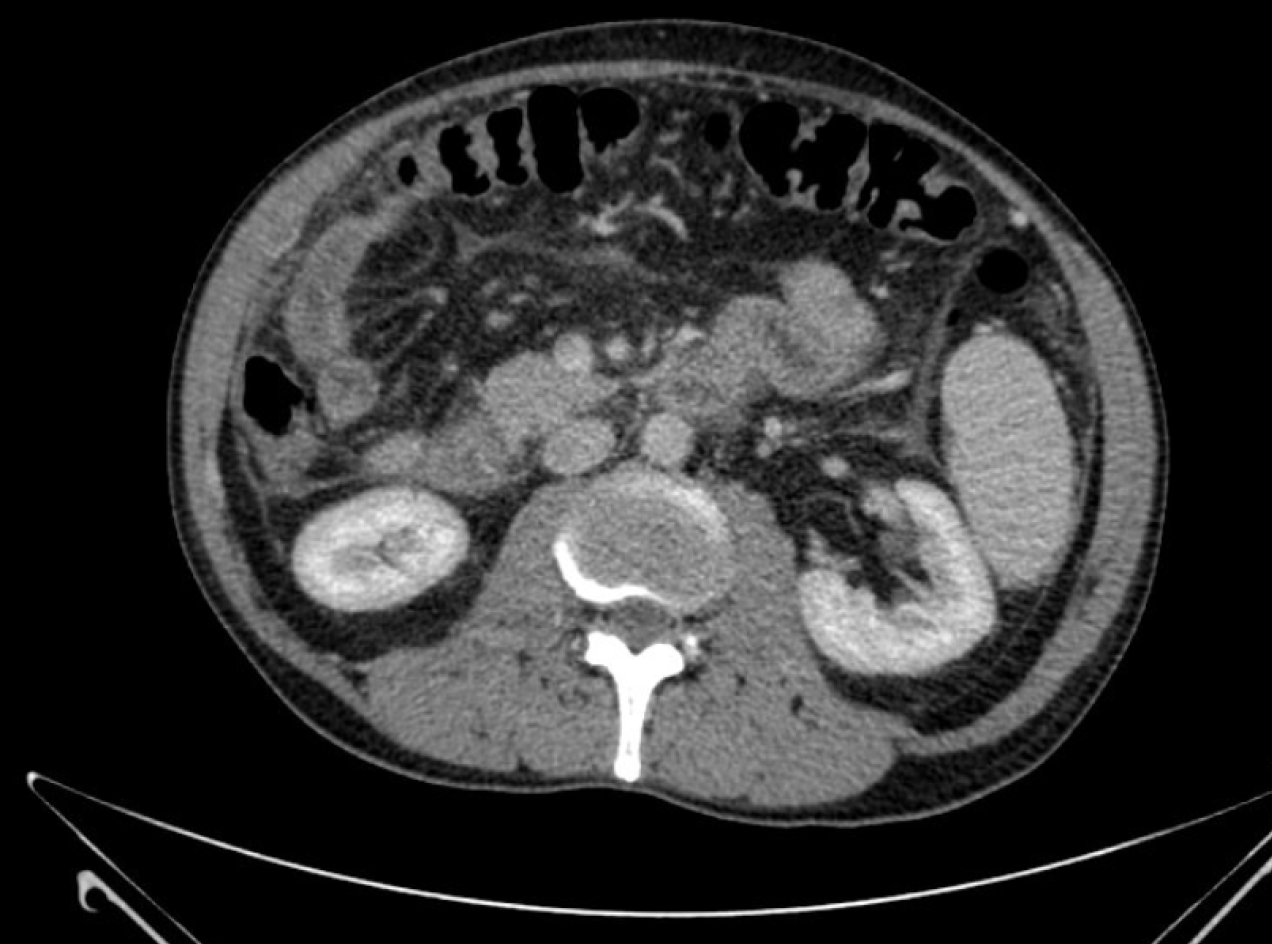
Figure 5 Computed tomography scan performed in September 2019.
Peritoneal carcinomatosis (before albumin-bound paclitaxel treatment).
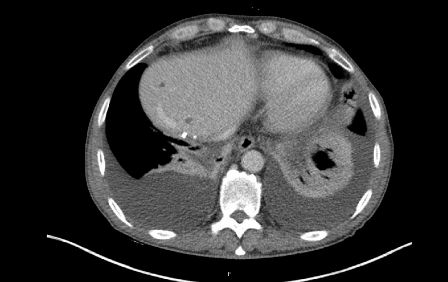
Figure 6 Computed tomography scan performed after four cycles of albumin-bound paclitaxel, where a mixed response was observed.
- Citation: Martin Huertas R, Fuentes-Mateos R, Serrano Domingo JJ, Corral de la Fuente E, Rodríguez-Garrote M. Albumin-bound paclitaxel as new treatment for metastatic cholangiocarcinoma: A case report. World J Clin Oncol 2020; 11(10): 844-853
- URL: https://www.wjgnet.com/2218-4333/full/v11/i10/844.htm
- DOI: https://dx.doi.org/10.5306/wjco.v11.i10.844


