Copyright
©The Author(s) 2019.
World J Clin Oncol. Apr 24, 2019; 10(4): 183-191
Published online Apr 24, 2019. doi: 10.5306/wjco.v10.i4.183
Published online Apr 24, 2019. doi: 10.5306/wjco.v10.i4.183
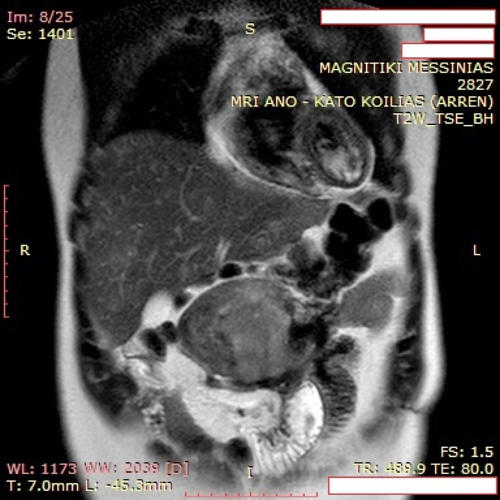
Figure 1 Magnetic resonance imaging revealed a mesenteric mass with heterogeneous intermediate and high signal intensity on T2-weighted images with parallel infiltration of surrounding small bowel loops.
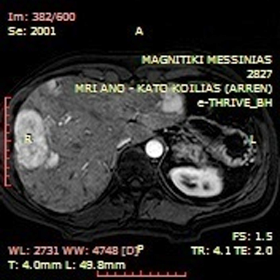
Figure 2 T1-weighted axial arterial phase post intravenous gadolinium administration image indicative of multiple hypervascular liver lesions compatible with focal nodular hyperplasia.
Figure 3 Desmoid type fibromatosis infiltrating the surrounding muscle fibers.
Eosin - hematoxylin, × 100 magnification.
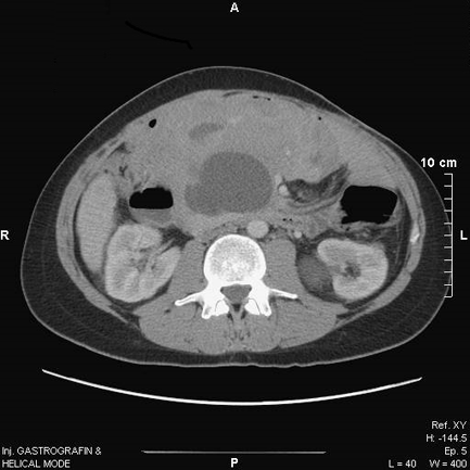
Figure 4 Computed tomography depiction of full disease response with liquification of the voluminous abdominal mass after one year of treatment with sorafenib.
- Citation: Mastoraki A, Schizas D, Vergadis C, Naar L, Strimpakos A, Vailas MG, Hasemaki N, Agrogiannis G, Liakakos T, Arkadopoulos N. Recurrent aggressive mesenteric desmoid tumor successfully treated with sorafenib: A case report and literature review. World J Clin Oncol 2019; 10(4): 183-191
- URL: https://www.wjgnet.com/2218-4333/full/v10/i4/183.htm
- DOI: https://dx.doi.org/10.5306/wjco.v10.i4.183


