Copyright
©The Author(s) 2019.
World J Clin Oncol. Nov 24, 2019; 10(11): 375-381
Published online Nov 24, 2019. doi: 10.5306/wjco.v10.i11.375
Published online Nov 24, 2019. doi: 10.5306/wjco.v10.i11.375
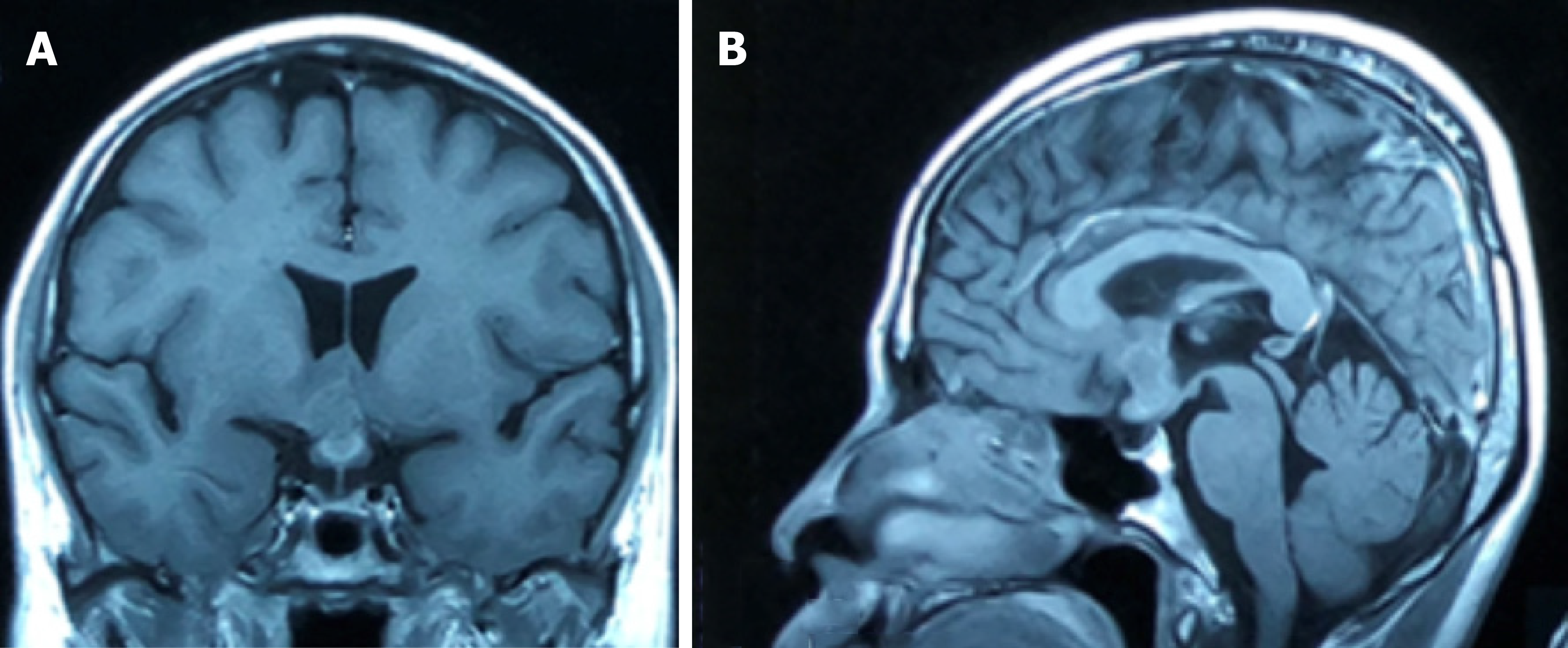
Figure 1 Magnetic resonance imaging of the brain, T1-weighted gadolinium-enhanced.
Coronal (A) and sagittal (B) view showing a heterogeneously enhanced lesion at the hypothalamic region with extension into the optic chiasm.
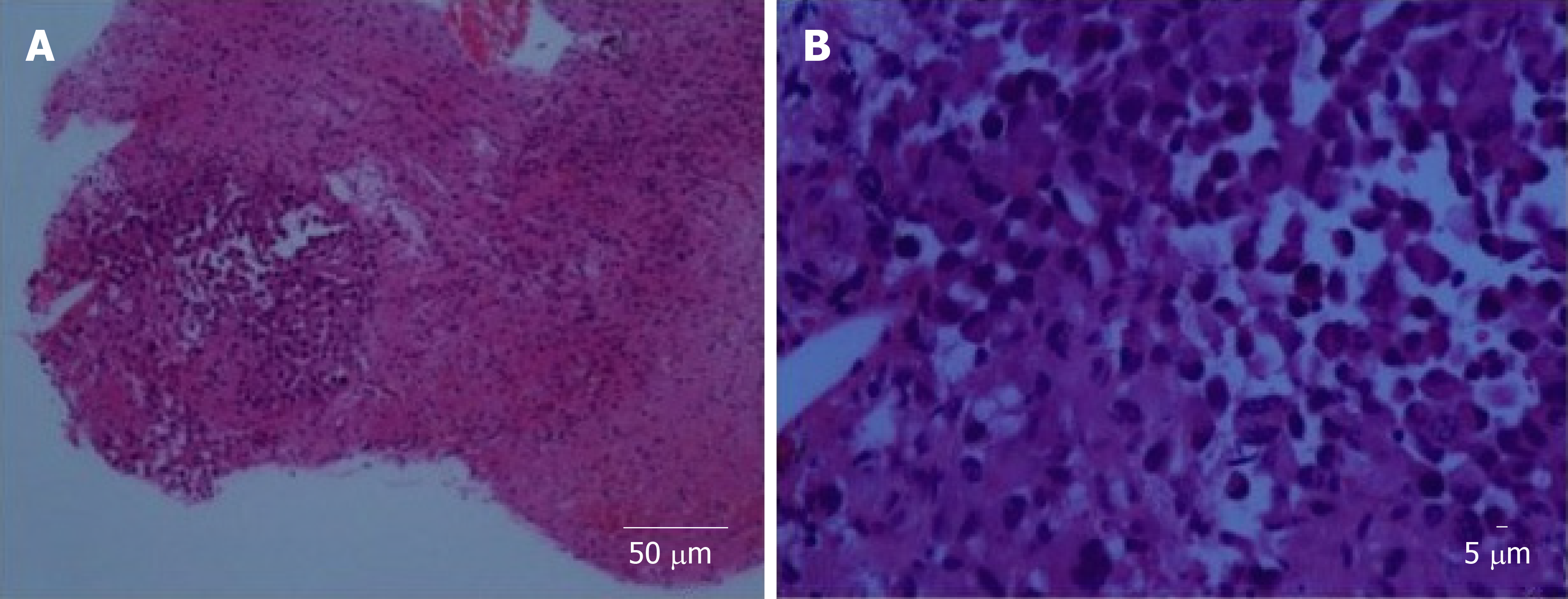
Figure 2 Histopathological examination.
A: Glial tissue infiltrated by a focus of singly dispersed malignant cells (40 ×); B: At high magnification, the lesion is composed of rhabdoid cells with hyper chromatic nuclei, coarse chromatin and abundant eosinophilic cytoplasm (400 ×).
- Citation: Ng PM, Low PH, Liew DNS, Wong ASH. Radiation-induced malignant rhabdoid tumour of the hypothalamus in an adult: A case report. World J Clin Oncol 2019; 10(11): 375-381
- URL: https://www.wjgnet.com/2218-4333/full/v10/i11/375.htm
- DOI: https://dx.doi.org/10.5306/wjco.v10.i11.375


