Copyright
©The Author(s) 2017.
World J Gastrointest Pathophysiol. May 15, 2017; 8(2): 87-92
Published online May 15, 2017. doi: 10.4291/wjgp.v8.i2.87
Published online May 15, 2017. doi: 10.4291/wjgp.v8.i2.87
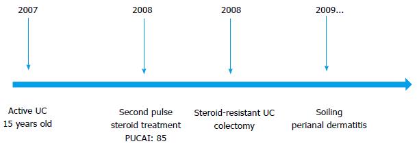
Figure 1 The medical treatment and follow-up of the patient.
UC: Ulcerative colitis.
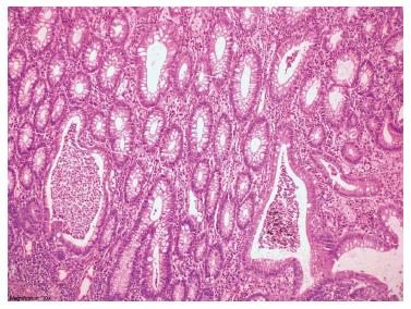
Figure 2 Histopathologic appearance of colonic mucosa.
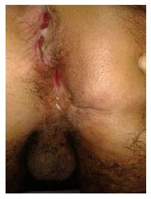
Figure 3 Perianal dermatitis and impaired perianal wound healing.
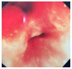
Figure 4 Two lumens visualized during ileoscopy.
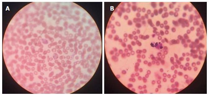
Figure 5 Nitroblue tetrazolium test.
A: Neutrophils from a patient with CGD fail to reduce the NBT dye and appear clear; B: Normal (unaffected) cells reduce the NBT dye and stain blue/purple. CGD: Chronic granulomatous disease; NBT: Nitroblue tetrazolium test.
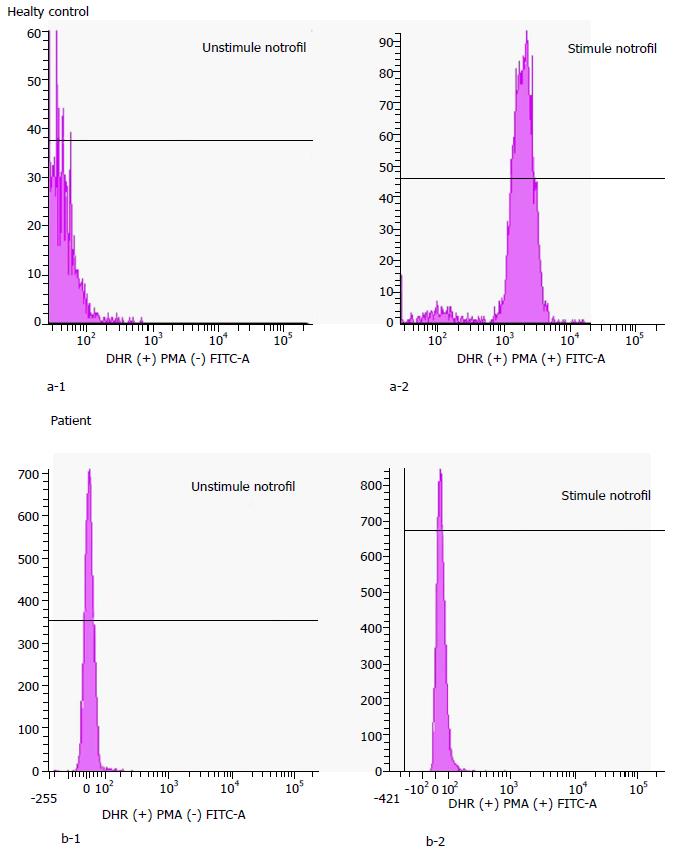
Figure 6 Flow cytometry-based dihydrorhodamine test.
DHR: Dihydrorhodamine.
- Citation: Kotlarz D, Egritas Gurkan O, Haskologlu ZS, Ekinci O, Aksu Unlusoy A, Gürcan Kaya N, Puchalka J, Klein C, Dalgic B. Differential diagnosis in ulcerative colitis in an adolescent: Chronic granulomatous disease needs extra attention. World J Gastrointest Pathophysiol 2017; 8(2): 87-92
- URL: https://www.wjgnet.com/2150-5330/full/v8/i2/87.htm
- DOI: https://dx.doi.org/10.4291/wjgp.v8.i2.87


