Copyright
©The Author(s) 2017.
World J Gastrointest Pathophysiol. May 15, 2017; 8(2): 59-66
Published online May 15, 2017. doi: 10.4291/wjgp.v8.i2.59
Published online May 15, 2017. doi: 10.4291/wjgp.v8.i2.59
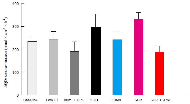
Figure 1 Differences in serosal vs mucosal oxygen consumption of rat sigmoid colon epithelium under several conditions.
No significant difference between groups was found, as assessed by one-way analysis of variance (P = 0.0849). ΔQO2: Serosa-mucosa, difference between serosal and mucosal oxygen consumption; Bum: Bumetanide; DPC: Diphenylamine-2-carboxylate; IBMX: 3-methyl-1-isobutilxanthine; SDR: Sodium-deprived rats; Ami: Amiloride.
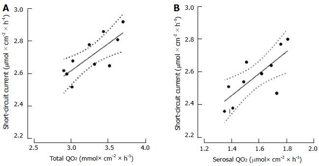
Figure 2 Linear regression of short-circuit current vs total oxygen consumption during normoxia (A), and vs serosal oxygen consumption during mucosal hypoxia (B).
Regression coefficients were 0.765 for A (P = 0.009) and 0.754 for B (P = 0.011). No outliers were detected.
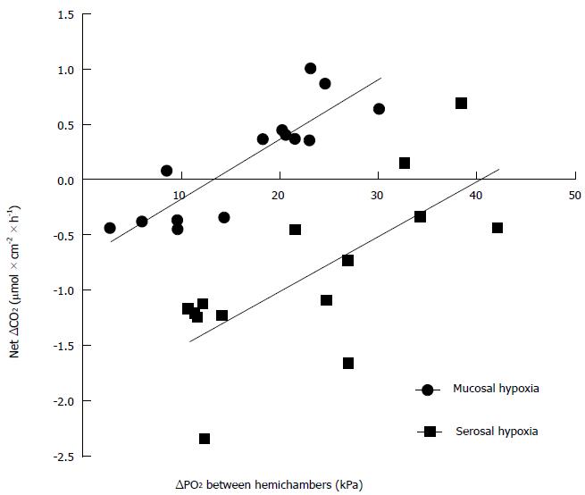
Figure 3 Rate of change in oxygen content of the hypoxic hemichamber as a function of the difference in oxygen partial pressure between hemichambers.
Various degrees of hypoxia were induced in either the serosal or the mucosal hemichamber while keeping the opposite hemichamber fully oxigenated, and the change in oxygen content was plotted as a function of the mean difference in oxygen pressure between both hemichambers during a 30-min observation period. The slope of the relationships was the same when either the serosal or the mucosal hemichamber was hypoxic (P = 0.8244), but the oxygen pressure difference at which there was no net change in oxygen content in the hypoxic hemichamber was larger when hypoxia was induced in the serosal hemichamber (P < 0.0001). ΔCO2: Change in oxygen content of the hypoxic hemichamber; ΔPO2: Oxygen pressure difference between hemichambers.
- Citation: Saraví FD, Carra GE, Matus DA, Ibáñez JE. Rectification of oxygen transfer through the rat colonic epithelium. World J Gastrointest Pathophysiol 2017; 8(2): 59-66
- URL: https://www.wjgnet.com/2150-5330/full/v8/i2/59.htm
- DOI: https://dx.doi.org/10.4291/wjgp.v8.i2.59


