Copyright
©2014 Baishideng Publishing Group Inc.
World J Gastrointest Pathophysiol. Aug 15, 2014; 5(3): 366-372
Published online Aug 15, 2014. doi: 10.4291/wjgp.v5.i3.366
Published online Aug 15, 2014. doi: 10.4291/wjgp.v5.i3.366
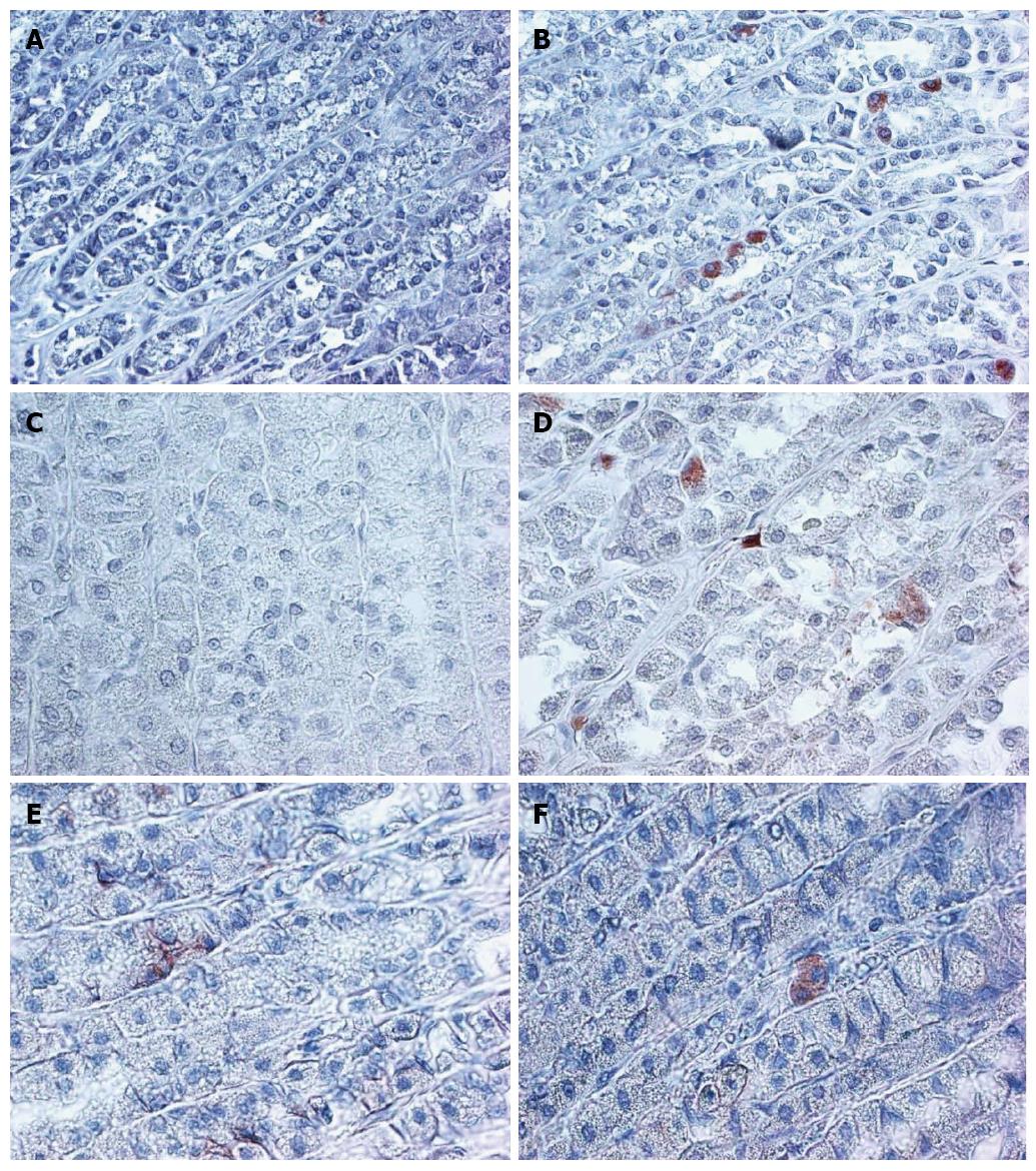
Figure 1 Higher expression of B-catenin, CD166, and LGR5 in normal aged rat gastric mucosa, compared to young rat normal gastric mucosa is demonstrated.
× 200 magnification. A: 4 m CD166; B: 24 m CD166; C: 4 m LGR5; D: 24 m LGR5; E: 4 m B-catenin; F: 24 m B-catenin.
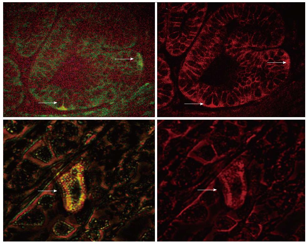
Figure 2 Double labeling for LGR5 (green, left panel) and B-catenin (red, right panel) in aged rat gastric mucosa (Lower Panel, × 600 magnification), H.
pylori infected human gastric mucosa (Upper Panel, × 400 magnification) shows cytoplasmic localization of B-catenin in an LGR5 expressing cell.
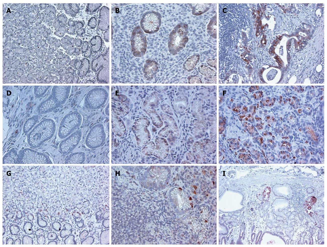
Figure 3 Immunohistochemical staining of stem cell markers ALDH1, CD166, LGR5 in human normal gastric mucosa, gastric mucosa with H.
pylori gastritis, and gastric adenocarcinoma, demonstrates increased expression of each of the markers over the normal controls. × 200 magnification. A: ALDH1 Normal; B: ALDH1 HP; C: ALDH1 CA; D: CD166 Normal; E: CD166 HP; F: CD166 CA; G: LGR5 Normal; H: LGR5 HP; I: LGR5 CA.
-
Citation: Levi E, Sochacki P, Khoury N, Patel BB, Majumdar AP. Cancer stem cells in
Helicobacter pylori infection and aging: Implications for gastric carcinogenesis. World J Gastrointest Pathophysiol 2014; 5(3): 366-372 - URL: https://www.wjgnet.com/2150-5330/full/v5/i3/366.htm
- DOI: https://dx.doi.org/10.4291/wjgp.v5.i3.366


