Copyright
©2010 Baishideng Publishing Group Co.
World J Gastrointest Pathophysiol. Dec 15, 2010; 1(5): 171-176
Published online Dec 15, 2010. doi: 10.4291/wjgp.v1.i5.171
Published online Dec 15, 2010. doi: 10.4291/wjgp.v1.i5.171
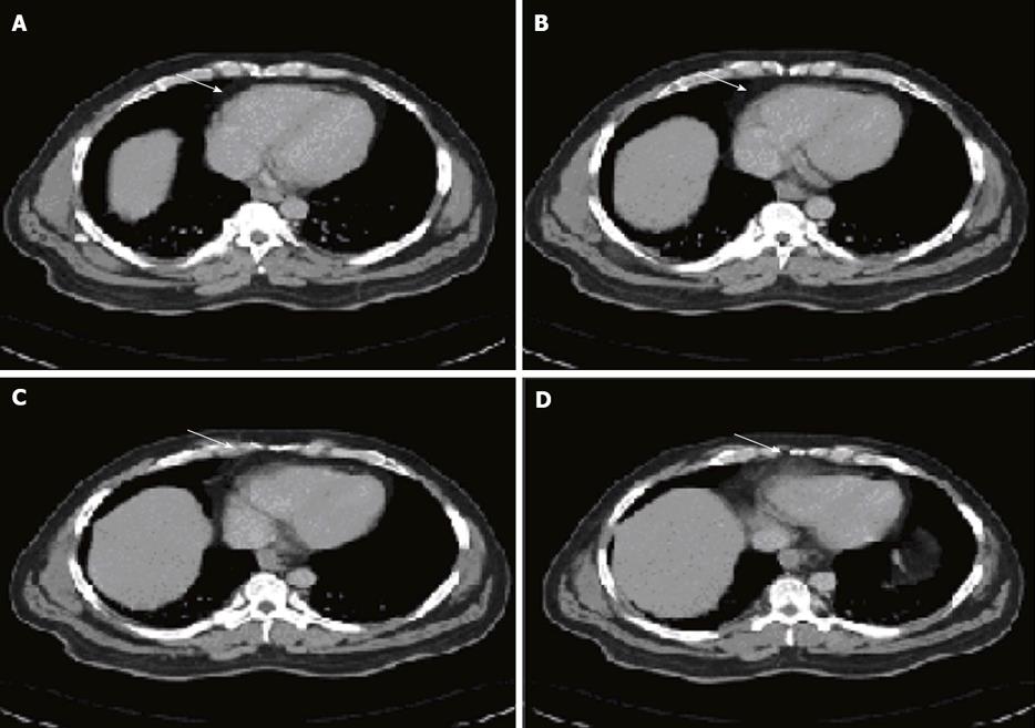
Figure 1 Abdominal computed tomography scan showing a large multilocular mass in the retroperitoneum (white arrows).
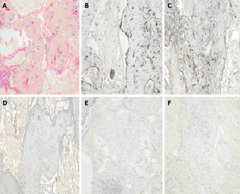
Figure 2 Photomicrograph of cystic lymphangioma.
A: Hematoxylin & Eosin staining demonstrating multiple dilated cystic spaces filled with pale pink proteinaceous material, fascicles of smooth muscles and inflammatory cells in stroma (× 100); B: CD31 immunostain showing positive staining of cystic lining endothelial cells (× 100); C: D2-40 antibody immunostain showing positive staining of cystic lining endothelial cells (× 100); D: Calretinin immunostain showing negative staining (× 100); E: HMB-45 immunostain showing negative staining (× 100); F: EMA immunostain showing negative staining (× 100).
- Citation: Bhavsar T, Saeed-Vafa D, Harbison S, Inniss S. Retroperitoneal cystic lymphangioma in an adult: A case report and review of the literature. World J Gastrointest Pathophysiol 2010; 1(5): 171-176
- URL: https://www.wjgnet.com/2150-5330/full/v1/i5/171.htm
- DOI: https://dx.doi.org/10.4291/wjgp.v1.i5.171


