Copyright
©2010 Baishideng Publishing Group Co.
World J Gastrointest Pathophysiol. Oct 15, 2010; 1(4): 144-146
Published online Oct 15, 2010. doi: 10.4291/wjgp.v1.i4.144
Published online Oct 15, 2010. doi: 10.4291/wjgp.v1.i4.144
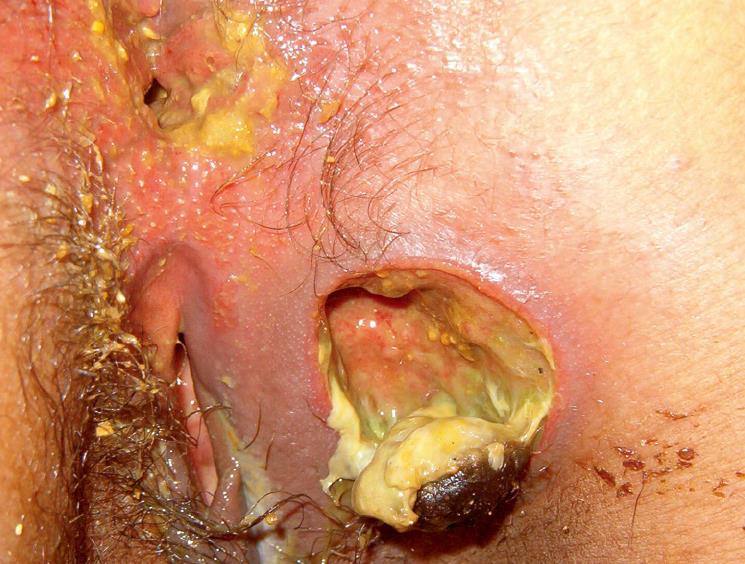
Figure 1 A rectal polypoid and ulcerated mass budding through the anal verge.
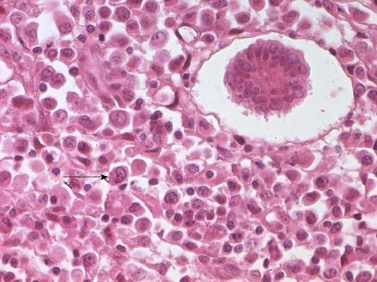
Figure 2 Tumoral cells with eosinophilic cytoplasm, round or convoluted nuclei with fine chromatin and one or more fine nucleolus (HE stain × 400).
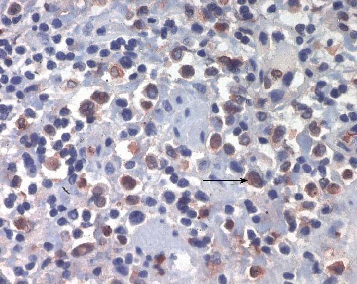
Figure 3 Cytoplasmic expression of myeloperoxydase (brown staining × 400).
- Citation: Benjazia E, Khalifa M, Benabdelkader A, Laatiri A, Braham A, Letaief A, Bahri F. Granulocytic sarcoma of the rectum: Report of one case that presented with rectal bleeding. World J Gastrointest Pathophysiol 2010; 1(4): 144-146
- URL: https://www.wjgnet.com/2150-5330/full/v1/i4/144.htm
- DOI: https://dx.doi.org/10.4291/wjgp.v1.i4.144


